Dogs can detect emotion in human voices, study shows

As many dog owners already know, dogs can often seem tuned into their “person’s” emotions or mood. Now, a new study from Current Biology offers evidence that this is, indeed, the case.

The study found that “dogs are sensitive to cues of emotion in human voices,”

And just how did researchers come to this conclusion? Fox reports, “For their study, researchers trained 11 dogs to sit still in a functional magnetic resonance imaging (fMRI) scanner. The researchers than analyzed the brain activity of both dogs and humans as they listened to 200 different dog and human sounds, ranging from crying to playful barking and laughing.

“While the brains of both dogs and humans responded most strongly to noises produced by their own species, they processed emotionally-loaded sounds in similar ways,” Fox reports.

“Some differences were noted as well: Dogs responded more strongly to non-vocal noises, compared to humans.”

Researchers hope the study will lead to “a better understanding of why dogs are so in tune with their owner’s emotions.”

Source: Now trending


Drops Are Best Treatment for Ear Tube-Related Dripping

For children with ear tubes, topical antibiotic drops treat the leaky discharge caused by an ear infection much more effectively than oral antibiotics or observation, according to a study published in The New England Journal of Medicine on Wednesday.

Each year, roughly 670,000 children in the United States have tiny plastic tubes placed in the eardrum in one of the most common surgeries of childhood. If the ear gets infected afterward, clear or bloody discharge can drip out, and a foul smell may be noticeable. This condition of drippy ears, which may or may not be painful, is known as tympanostomy tube otorrhea. In a 2013 study, 67 percent of children who had ear tubes put in experienced one or more episodes of otorrhea in the year after the procedure.

Smaller trials with different designs have found that ear drops are more effective than systemic antibiotics for this common problem. But the new study is the first to include a no-treatment, wait-and-see group, and provides the best evidence to date for the superiority of ear drops for children with tubes.

“This is a big study, very high quality and very rigorous. It’s more of a definitive study,” said Dr. Richard M. Rosenfeld, chairman of otolaryngology at SUNY Downstate Medical Center in Brooklyn, who was not involved in the research. Putting drops into the ear canal, he said, is akin to “dropping a Scud missile on the bacteria.”

There are two benefits, he said. “It resolves the otorrhea more effectively and faster than oral medicine,” he said. “More importantly, you avoid the problem of resistant germs, which is a major, major problem.”

In the new study, 230 children ages 1 to 10 with acute tube discharge were randomly assigned to three groups: some got ear drops, some got oral antibiotics, and some were simply observed.

At two weeks, 5 percent of the children treated with drops still had discharge from an infection, compared with 44 percent of those treated with oral antibiotics and 55 percent of those who were only observed.

The study suggests that drippy ears in children with tubes might take two weeks or longer to resolve without treatment — a long time if the child has trouble sleeping or cannot participate in activities.

“No previous study assessed the actual need to treat these children,” said Dr. Thijs M.A. van Dongen, the lead author of the study and an epidemiologist at University Medical Center Utrecht in the Netherlands.

In a six-month follow-up, the study found that children who were not treated for the first two weeks had a median total of 18 days of discharge, compared with five days for those who got ear drops, and 13.5 days for those given oral antibiotics.

“It’s better to start treatment quickly after onset of symptoms,” Dr. van Dongen said. “They improve more quickly, and they have less recurrence in following months.”

But Dr. Rosenfeld said it was not clear that all children with tube otorrhea should promptly start ear drops. Watching and waiting is an option, he said, if the drainage is not profuse and causes no discomfort, and the child still sleeps and acts normally. Drops can be expensive, and if used excessively they can cause hard-to-treat yeast overgrowth.

Last July, practice guidelines issued by the American Academy of Otolaryngology-Head and Neck Surgery strongly recommended that clinicians prescribe only topical antibiotic ear drops for children with uncomplicated cases of tube discharge.

But some pediatricians and family physicians still routinely prescribe systemic antibiotics for these cases. Among otolaryngologists, the new guidelines are “fairly well accepted,” said Dr. Joseph E. Kerschner, a professor of otolaryngology and the dean of the medical school at Medical College of Wisconsin in Milwaukee. “Still, there’s evidence that not all physicians have gotten the message.”

A 2013 survey found that 54 percent of emergency-medicine physicians used oral antibiotics to treat an ear infection in a child with ear tubes, compared with just 9 percent of ear, nose and throat doctors, almost all of whom used topical antibiotics.

Most children do not see ear, nose and throat specialists first, said Dr. Seth R. Schwartz, an otolaryngologist and the director of the Listen for Life Center at Virginia Mason Medical Center in Seattle, so “it’s important that all physicians who treat children with this condition are aware.”

The bottom line is that “oral antibiotics don’t work well” in these cases, he said, and they may cause stomach upset or diarrhea.

In an uncomplicated case of tube discharge, Dr. Kerschner advised parents to say to pediatricians, “’Hey, my kid has a draining ear, how about using topical antibiotics instead of oral antibiotics?’ The child will get better faster and more reliably.”

In children without ear tubes who get an ear infection, drops are not effective as they cannot get behind the ear drum. For those children, oral antibiotics are a common treatment. Lately, Dr. Schwartz said, “there’s a higher recognition that you can treat with observation initially with close follow-up.”

Source: New York Times


Prostate’s Early Growth May Reveal Cures for Later Illnesses

Dr. David Samadi is the chairman of urology and chief of robotic surgery at Lenox Hill Hospital in New York City and is a board-certified urologist and oncologist specializing in the diagnosis and treatment of urologic diseases, kidney cancer, bladder cancer and prostate cancer. Samadi also specializes in many advanced, minimally invasive treatments for prostate cancer; is one of the few urologic surgeons in the United States trained in oncology, open-, laparoscopic- and robotic-surgery; and was the first surgeon in the nation to successfully perform a robotic surgery redo. He contributed this article to Live Science’s Expert Voices: Op-Ed & Insights.

For a surgeon who has successfully treated prostate cancer in many thousands of men by removing their prostate gland, the idea that science might one day be able to regenerate this gland using stem cells is a foreign one — and yet highly intriguing. But this advancement is just one of many potential treatments for prostate cancer or benign prostate enlargement that may eventually arise from important new research on the cellular building blocks of prostate gland development.

In a study published Feb. 11 in the journal Stem Cell Reports, scientists from the University of York in England detailed their discovery of a “signaling pathway,” a set of signals that tell proteins inside stem cells how to evolve into prostate tissue cells called basal cells and luminal cells. The researchers learned there are 80 genes involved in this process, and that the main signals responsible for activating prostate development are retinoic acid and male sex hormones — the balance of which are disrupted in prostate cancer.

Source: live science


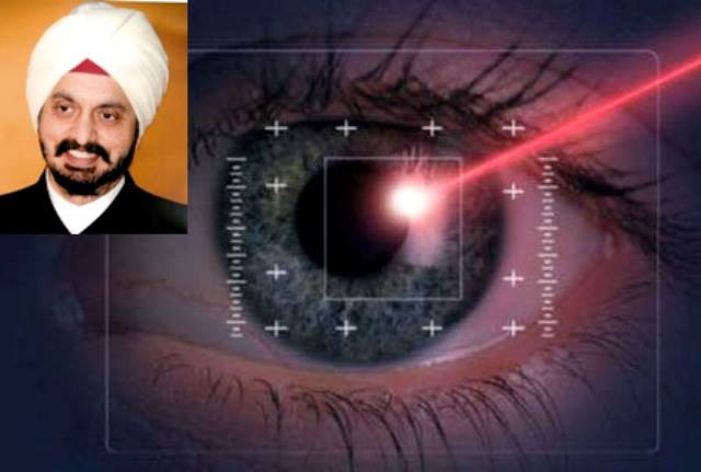
New layer of cornea discovered by Indian doctor Harminder Dua

A new layer in the human cornea was discovered by the Indian doctor Harminder Dua at The University of Nottingham in 2014. It plays a vital role in the structure of the tissue that controls the flow of fluid from the eye.

The research was published in a paper in the British Journal of Ophthalmology.
The new finding could shed new light on glaucoma. The new layer named as Dua’s Layer is just 15 microns thick but incredibly tough. Comprised of thin plates of collagen, it sits at the back of the cornea between the corneal stroma and Descemet’s membrane.

It makes an important contribution to the sieve-like meshwork, the trabecular meshwork (TM), in the periphery of the cornea.

The TM is a wedge-shaped band of tissue that extends along the circumference of the angle of the anterior chamber of the eye.

It is made of beams of collagen wrapped in a basement membrane to which trabecular cells and endothelial cells attach. The beams branch out randomly to form a ‘meshwork’.
Scientists had previously believed the cornea to be comprised of five layers – from front to back— the corneal epithelium, Bowman’s layer, the corneal stroma, Descemet’s membrane and the corneal endothelium.

Pressure within the eye is maintained by the balance of aqueous fluid production by eye tissue called the ciliary body and drainage principally through the TM to the canal of Schlemm, a circular channel in the angle of the eye.

Defective drainage through the TM is an important cause of glaucoma, a condition that leads to raised pressure in the eye that can permanently affect sight. Around 1 to 2% of the world’s population yearly have chronic glaucoma and globally around 45 million people have open angle glaucoma which can permanently damage the optic nerve – 10% of whom are blind.

It is hoped the discovery will offer new clues on why the drainage system malfunctions in the eyes of some people, leading to high pressure.
Glaucoma is a devastating disease caused by defective drainage of fluid from the eye. Glaucoma is the second largest leading cause of blindness of world.

Source: Zee News


Pain ‘dimmer switch’ discovered by UK scientists

Pain sensitivity is controlled by a genetic “dimmer switch”, which can be re-set, UK scientists have discovered.

Twins sharing 100% of genes have different pain thresholds, which can potentially be altered by lifestyle or medication, say researchers at King’s College, London.

The study could lead to new painkillers or lifestyle interventions, they report in Nature Communications.

One in five of the population suffers from acute or chronic pain.

Lead researcher Dr Jordana Bell said the potential to regulate genes involved in pain sensitivity “is very exciting and could lead to a more effective pain relief treatment for patients suffering with chronic pain”.

Sensitivity to pain is complex, with wide individual variation. Previous studies have suggested about half of the influence is explained by genes.

To identify levels of sensitivity to pain, scientists tested 25 pairs of identical twins using a heat probe placed on the arm.

Identical twins share 100% of their genes; therefore any difference between identical twins must be due to their environment or changes affecting the function of their genes.

Study participants were asked to press a button when the heat became painful for them, which allowed the researchers to determine their pain thresholds.

Using DNA sequencing, the researchers examined the whole genetic codes (genomes) of the twins and compared them with 50 unrelated individuals.

The research team found chemical changes within nine genes involved in pain sensitivity that were different in one twin but not in her identical sister.

These were most significant within a known pain sensitivity gene, which is already a target for the development of new painkillers.

Research into the switching on and off of genes, a process known as epigenetic regulation, is a big growth area for the development of new medicines.

‘Landmark’ study
Tim Spector, professor of genetic epidemiology at King’s College London, said epigenetic switching is “like a dimmer switch for gene expression”.

“This landmark study shows how identical twins, when combined with the latest technology to look at millions of epigenetic signals, can be used to find the small chemical switches in our genes that make us all unique – and in this case respond to pain differently.”

The chemical changes act like a “thermostat” or “dimmer switch” to set an individual’s pain sensitivity, Prof Spector added.

“Using drugs or changes in lifestyle, we might be able to reset that thermostat, allowing that person in the future to feel less pain,” he told BBC News.

“The epigenetic changes are potentially reversible.”

Source: BBC news


Cold weather can help you lose weight, study says

Keeping temperatures a bit chillier at home and work, even when it’s as cold as it is now, can make your body burn more calories to keep warm, Dutch researchers say.

Americans love to crank up the thermostat, especially in bitterly cold times like these.

But a new study suggests turning it down a few degrees could actually help you lose weight.

We know. Not what you wanted to hear right now.

But Dutch researchers say regular exposure to mildly cold temperatures can make your body burn more calories to keep warm.

“Since most of us are exposed to indoor conditions 90 percent of the time, it is worth exploring health aspects of ambient temperatures,”. “What would it mean if we let our bodies work again to control body temperature?”

Lichtenbelt and his team have been studying the phenomena for the past 10 years.

While most animals (humans included) shiver to stay warm, another type of shivering — called non-shivering thermogenesis — occurs when the temperature is cool but not cold, according to the research.

That type of shivering, activating what’s called “brown fat,” can burn up to 30 percent of the body’s energy and contribute to weight loss.

Brown fat, discovered in adults in 2009, burns calories instead of storing them like white fat.

So does this mean you should crank the heat down to 55 degrees and frolic about in a tank top and underwear?

Not necessarily. It’s more theory at this point, but researchers also said it wouldn’t hurt.

“It would do no harm,” Dr. Mitchell Lazar, chief of the division of endocrinology, diabetes and metabolism at the University of Pennsylvania, told HealthDay. “It’s worth a try for someone who is having trouble losing weight by diet and exercise alone.”

Source: global post


Will seeing red help you lose weight?

Previously, scientists found diners at a pasta buffet heaped the marinara on if they used white plates, but took smaller helpings if their plates were red. They did the opposite when the pasta had a white sauce. So researchers thought the key to eating less might be sharp color contrasts.

But the new study, published in the journal Appetite this month, indicates it’s not contrast, but one specific color — red — that causes people to cut back on what they consume. The research tested how much food or hand cream people used when the product was placed on a red, white or blue plate.

“We wanted to find out if the effect was limited to eating or generalized to other types of consumption. The cream was a convenient way to evaluate another sensory system — touch, rather than taste,” said study author Nicola Bruno, cognitive psychology researcher at the University of Parma, Italy.

The study
In the new study, volunteers rated the saltiness of popcorn, nuttiness of chocolate and stickiness of hand cream.

Each person received a pre-measured sample of a product on a plate that was one of three colors — red, white or blue. The volunteers munched and moisturized as much as they liked while they filled out their answers. Of the 240 participants, 90 taste-tested popcorn, and 75 each sampled the chocolate chips and hand cream.

Each survey also included a question to check how much testers liked the product, since this may have triggered them to eat or use more. After the experiments, researchers measured how much the testers had consumed.

The authors also measured differences in the color intensity and contrasts of foods, cream and plates. Data in hand, they tested whether differences in people’s consumption correlated with differences in color contrast.

Results
On average, people ate less popcorn and chocolate when they were served on red plates compared to blue or white plates.

Not surprisingly, self-reported popcorn fans ate more than those who expressed no preference for it on the survey. However, these people consumed more kernels independent of plate color. When researchers corrected for people’s preferences in their statistical analysis, eating off red plates was still associated with lower consumption.
Use of the moisturizing cream followed a similar trend. When testing hand cream on red plates, people used about half as much, on average, compared to cream on blue or white plates.

Contrast had little to do with these results, said Bruno. Though dark chocolate on a red plate offered less contrast than pale colored popcorn or cream, people still took fewer chocolate chips.

“I expected to find the results related to differences in color intensity, but they did not. It’s really related to the color red compared to the food and cream colors,” he said.

Limitations
The study supports the idea that the color red reduces consumption, according to Oliver Genschow, who studies consumer psychology at the University of Mannheim.
But don’t run out and buy those red plates as a holiday gift just yet. In all the research so far, participants were unaware of the real reason for the tests, implying an unconscious process may be at work.

“We don’t know what will happen if people are conscious of their plate’s color. Maybe it won’t work anymore,” Genschow said.

He says color may be an additional factor to consider when treating patients with certain eating disorders, but it’s premature to suggest everyone trying to lose weight should simply switch to red plates.

Source: cnn news


Neurons in spinal cord send Cc of commands back to brain

Research group led by Professor Silvia Arber at the University of Basel’s Biozentrum and the Friedrich Miescher Institute for Biomedical Research has now discovered, that many neurons in the spinal cord send their instructions not only towards the musculature, but at the same time also back to the brain via an exquisitely organized network.
This dual information stream provides the neural basis for accurate control of arm and hand movements.

Movements of our arms and hands, in particular, call for extremely precise coordination.
The brain sends a constant stream of commands via the spinal cord to our muscles to execute a wide variety of movements.

This stream of information from the brain reaches interneurons in the spinal cord, which then transmit the commands via further circuits to motor neurons innervating muscles.

The research group led by Silvia Arber at the Biozentrum of the University of Basel and the Friedrich Miescher Institute for Biomedical Research has now elucidated the organization of a second information pathway taken by these commands.
The scientists showed that many interneurons in the mouse spinal cord not only transmit their signals via motor neurons to the target muscle, but also simultaneously send a copy of this information back to the brain.

“The motor command to the muscle is sent in two different directions – in one direction, to trigger the desired muscular contraction and in the other, to inform the brain that the command has actually been passed on to the musculature,” Chiara Pivetta, first author of the publication, said.

In analogy to e mail transmission, the information is thus not only sent to the recipient but also to the original requester.
What happens to the information sent by spinal interneurons to the brain? As Arber’s group discovered, this input is segregated by function and spatially organized within a brainstem nucleus.

Information from different types of interneurons thus flows to different areas of the nucleus. For example, spinal information that will influence left-right coordination of a movement is collected at a different site than information affecting the speed of a movement.

The findings are published in the journal Cell.

Source: Yahoo news


Peanut allergy treatment ‘a success’

Doctors say a potential treatment for peanut allergy has transformed the lives of children taking part in a large clinical trial.

The 85 children had to eat peanut protein every day – initially in small doses, but ramped up during the study.

The findings, published in the Lancet, suggest 84% of allergic children could eat the equivalent of five peanuts a day after six months.

Experts have warned that the therapy is not yet ready for widespread use.

Peanuts are the most common cause of fatal allergic reactions to food.

There is no treatment so the only option for patients is to avoid them completely, leading to a lifetime of checking every food label before a meal.

The trial, at Addenbrooke’s Hospital in Cambridge, tried to train the children’s immune systems to tolerate peanut protein.

Every day they were given a peanut protein powder – starting off on a dose equivalent to one 70th of a peanut.

The theory was that patients started at the extremely low dose, well below the threshold for an allergic response.

Once a fortnight the dose was increased while the children were in hospital, in case there was an reaction, and then they continued taking the higher dose at home.

The majority of patients learned to tolerate the peanut.

Lena Barden, 11, from Histon in Cambridgeshire, said: “It meant a trip to the hospital every two weeks.

“A year later I could eat five whole peanuts with no reaction at all.

“The trial has been an experience and adventure that has changed my life and I’ve had so much fun, but I still hate peanuts!”

‘Dramatic transformation’
One of the researchers, Dr Andrew Clark, told the BBC: “It really transformed their lives dramatically; this really comes across during the trial.

“It’s a potential treatment and the next step is to make it available to patients, but there will be significant costs in providing the treatment – in the specialist centres and staff and producing the peanut to a sufficiently high standard.”

Fellow researcher Dr Pamela Ewan added: “This large study is the first of its kind in the world to have had such a positive outcome, and is an important advance in peanut allergy research.”

But she said further studies would be needed and that people should not try this on their own as this “should only be done by medical professionals in specialist settings”.

The research has been broadly welcomed by other researchers in the field, but some concerns about how any therapy could be introduced have been raised.

Caution
Prof Gideon Lack, who is running a peanut allergy trial at the Evelina Children’s Hospital in London, told the BBC: “This is a really important research step in trying to improve our management of peanut allergy, but is not yet ready for use in clinical practice.

“We need a proper risk assessment needs to be done to ensure we will not make life more dangerous for these children.

He warned that 60% of people with a peanut allergy were also allergic to other nuts so a carefree lifestyle would rarely be an option.

Prof Barry Kay, from the department of allergy and clinical immunology at Imperial College London, said: “The real issues that still remain include how long the results will last, and whether the positive effects might lead affected individuals to have a false sense of security.

“Another issue to address is whether there will be long term side-effects of repeated peanut exposure even where full allergic reaction does not occur, such as inflammation of the oesophagus.

“So, this study shows encouraging results that add to the current literature, but more studies are needed to pin down these issues before the current advice to peanut allergy sufferers, which is to avoid eating peanuts, is changed.”

Maureen Jenkins, director of clinical services at Allergy UK, said: “The fantastic results of this study exceed expectation.

“Peanut allergy is a particularly frightening food allergy, causing constant anxiety of a reaction from peanut traces.

Source: BBC news


New method makes stem cells in about 30 minutes, scientists report

In a feat that experts say is a significant advance for regenerative medicine, scientists have discovered a surprisingly simple method for creating personalized stem cells that doesn’t involve human embryos or tinkering with DNA.

Two studies published Wednesday in the journal Nature describe a novel procedure for “reprogramming” the blood cells of newborn mice by soaking the cells in a mildly acidic solution for 30 minutes. This near-fatal shock caused the cells to become pluripotent, or capable of growing into any type of cell in the body.

When the reprogrammed cells were tagged and injected into a developing mouse, they multiplied and grew into heart, bone, brain and other organs, the scientists found.
“It was really surprising to see that such a remarkable transformation could be triggered simply by stimuli from outside of the cell,” said lead study author Haruko Obokata, a biochemistry researcher at the RIKEN research institute in Japan. “Very surprising.”

The simplicity of the technique, which Obokata and her colleagues dubbed stimulus triggered acquisition of pluripotency, or STAP, caught many experts off-guard.
“So you mistreat cells under the right conditions and they assume a different state of differentiation? It’s remarkable,” said Rudolf Jaenisch, a pioneering stem cell researcher at MIT who was not involved in the study. “Let’s see whether it works in human cells, and there’s no reason why it shouldn’t.”

Obokata said that researchers had already begun experiments on human cells, but offered no details.

Due to their Zelig-like ability to form any number of specialized cells, pluripotent stem cells are considered the basic building blocks of biology. Scientists are working on ways to use them to repair severed spinal cords, replace diseased organs, and treat conditions as varied as diabetes, blindness and muscular dystrophy.

By using stem cells spawned from the patient’s own cells, replacement tissues would stand less of a chance of being attacked by the patient’s own immune system, researchers say. That would spare patients the need to undergo a lifetime regimen of dangerous, immune-suppressing drugs.

But progress toward these lofty goals has been slow, due in part to the challenges of current stem cell production methods. The practice of harvesting stem cells from human embryos makes many people uncomfortable, and some religious groups have pressed for limits or bans on their use. Even scientists who want to study them say they may not be practical for medical therapies because they could be rejected by a patient’s body.

Another approach is to rewind a patient’s own mature cells to a pluripotent state. Dr. Shinya Yamanaka, the first person to make these induced pluripotent stem cells, won a Nobel Prize for this work in 2012. However, the reprogramming process converts only about 1% of the cells into iPS cells, and questions remain about their long-term stability and safety.

The STAP method presents a simpler, cheaper and faster method of producing stem cells, said Chris Mason, a professor of regenerative medicine bioprocessing at University College London.

“How much easier can it possibly get,” Mason told the Science Media Centre, an English organization that promotes scientific understanding on controversial subjects.
“If it works in man, this could be the game changer that ultimately makes a wide range of cell therapies available using the patient’s own cells as starting material,” he said. “The age of personalized medicine would have finally arrived.”

The STAP approach was inspired by observations of plant cells that changed character when they were exposed to environmental stress, according to the research team from RIKEN and Harvard’s Brigham and Women’s Hospital in Boston.

Obokata and her colleagues set about “stressing” mouse blood cells in a variety of ways to see if they would change. They exposed them to heat, deprived them of nutrition and repeatedly poured them through narrow glass pipes.

The method they ultimately published involved placing the cells in an acid solution for 30 minutes and then spinning them in a centrifuge for five minutes. The process converted 7% to 9% of the original cells into STAP cells, Obokata said.

To see whether the cells had been reprogrammed, researchers engineered the mice with a gene that would cause their cells to glow a fluorescent green under ultraviolet light if they became pluripotent. After torturing the blood cells, they began to glow after three days and appeared to peak at seven days, suggesting that they had become pluripotent in just a week’s time. The researchers bolstered the cells’ ability to proliferate by treating them with hormones and an immune cell secretion called leukemia inhibitory factor.

To fully prove that they had become pluripotent, the STAP cells were injected into normal mouse embryos. The resulting offspring, called a chimera, were a mix of regular cells and glowing STAP cells.

Andrew McMahon, director of USC’s Eli and Edyth Broad Center for Regenerative Medicine, said the creation of a chimera was critical to proving that blood cells had changed in a fundamental way.

“That’s the most rigorous [test] you could possibly do,” said McMahon, who was not involved in the study. It shows that the STAP cells can make every type of cell in the embryo and that they “can organize in a normal-looking way, so that what comes out is a normal looking fetus.”

McMahon said the study was also surprising in that it showed that mature cells could be reprogrammed without having to divide.

“That’s why the change is so rapid, because the cells don’t have to undergo division for this to occur,” he said. “It’s a really interesting and novel finding.”

Yamanaka, who was not involved in the STAP study, said the research would undoubtedly help scientists understand the basic biology of cellular reprogramming.

“The findings are important,” said Yamanaka, who directs Kyoto University’s Center for iPS Cell Research.

The reasons why stress causes cells to drastically alter their function remains a mystery, Obokata and her colleagues said.

She declined to say whether the researchers were seeking a patent on the STAP procedure.

Source: latimes