Useful safety tips for epilepsy patients

Safety in the home starts with your kitchen and your bathrooms. These are the places where, during and after any seizure, you can become confused and risk injury. Take these seizure precautions to decrease the chance of accidents.

Safeguard your kitchen

  • Use oven mitts and cook only on rear burners
  • If possible, use an electric stove, so there is no open flame
  • Cooking in a microwave is the safest option
  • Ask your plumber to install a heat-control device in your faucet so the water doesn’t become too hot
  • Carpet the kitchen floor. This can provide cushioning if you fall
  • Use plastic containers rather than glass when possible

Safeguard your bathroom

  • Install a device in your tub and showerhead that controls temperature. This keeps you from burning yourself if a seizure occurs
  • Carpet the floor—it’s softer and less slippery than tile
  • Do not put a lock on the bathroom door. If you have one, never use it. Someone should always be able to get in if you need help
  • Learn to bathe with only a few inches of water in the tub, or use a handheld showerhead
  • Planning ahead for safety outside the home

Driving.

For many people with epilepsy, the risk of seizures restricts their independence, in particular the ability to drive. The Epilepsy Foundation offers a state-by-state database of driving restrictions and regulations on its website. Find out more about driving and epilepsy.
Participating in activities.

You can play sports with epilepsy, but it’s a good idea to have someone with you who knows how to manage a seizure. Wearing head protection is also recommended when you participate in a contact sport that might cause you to fall or hit your head.

Here are some tips for picking the right physical activities when you are living with epilepsy:

  • If seizures usually occur at a certain time, plan activities when seizures are less likely to happen
  • Avoid extreme heat when exercising and keep hydrated with plenty of water to reduce seizure risks
  • Check with your neurologist before starting any new exercise program

Some activities may be restricted if you have uncontrolled seizures, including:

  • Swimming alone
  • Climbing to unsafe heights
  • Riding a bike in traffic

Source: VIMPAT


Neurons in spinal cord send Cc of commands back to brain

Research group led by Professor Silvia Arber at the University of Basel’s Biozentrum and the Friedrich Miescher Institute for Biomedical Research has now discovered, that many neurons in the spinal cord send their instructions not only towards the musculature, but at the same time also back to the brain via an exquisitely organized network.
This dual information stream provides the neural basis for accurate control of arm and hand movements.

Movements of our arms and hands, in particular, call for extremely precise coordination.
The brain sends a constant stream of commands via the spinal cord to our muscles to execute a wide variety of movements.

This stream of information from the brain reaches interneurons in the spinal cord, which then transmit the commands via further circuits to motor neurons innervating muscles.

The research group led by Silvia Arber at the Biozentrum of the University of Basel and the Friedrich Miescher Institute for Biomedical Research has now elucidated the organization of a second information pathway taken by these commands.
The scientists showed that many interneurons in the mouse spinal cord not only transmit their signals via motor neurons to the target muscle, but also simultaneously send a copy of this information back to the brain.

“The motor command to the muscle is sent in two different directions – in one direction, to trigger the desired muscular contraction and in the other, to inform the brain that the command has actually been passed on to the musculature,” Chiara Pivetta, first author of the publication, said.

In analogy to e mail transmission, the information is thus not only sent to the recipient but also to the original requester.
What happens to the information sent by spinal interneurons to the brain? As Arber’s group discovered, this input is segregated by function and spatially organized within a brainstem nucleus.

Information from different types of interneurons thus flows to different areas of the nucleus. For example, spinal information that will influence left-right coordination of a movement is collected at a different site than information affecting the speed of a movement.

The findings are published in the journal Cell.

Source: Yahoo news


New drug target for Alzheimer’s identified

Researchers have identified abnormal expression of genes, resulting from DNA relaxation, that can be detected in the brain and blood of Alzheimer’s patients.
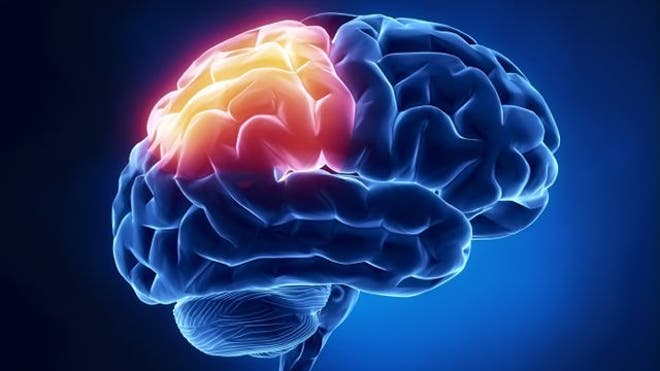
The protein tau is involved in a number of neurodegenerative disorders, including Alzheimer’s disease.

Previous studies have implicated DNA damage as a cause of neuron, or cell, death in Alzheimer’s patients.

Given that DNA damage can change the structure of DNA within cells, the researchers led by Bess Frost examined changes in DNA structure in tau-induced neurodegeneration.

They used transgenic flies and mice expressing human tau to show that DNA is more relaxed in tauopathy.

They then identified that the relaxation of tightly wound DNA and resulting abnormal gene expression are central events that cause neurons to die in Alzheimer’s disease.

“Our work suggests that drugs that modify DNA structure may be beneficial for treating Alzheimer’s Disease,” they wrote.

The study is published in the journal Nature Neuroscience.

Source: Business Standard


Childhood amnesia occurs at the age of seven

Psychologists have suggested that age seven is when these earliest memories tend to fade into oblivion, a phenomenon known as “childhood amnesia.”

The research involved interviewing children about past events in their lives, starting at age three. Different subsets of the group of children were then tested for recall of these events at ages five, six, seven, eight and nine.

Emory psychologist Patricia Bauer, who led the study, said that their study is the first empirical demonstration of the onset of childhood amnesia.

She said that they actually recorded the memories of children, and then they followed them into the future to track when they forgot these memories.

The experiment began by recording 83 children at the age of three, while their mothers or fathers asked them about six events that the children had experienced in recent months, such as a trip to the zoo or a birthday party.

After recording these base memories, the researchers followed up with the children years later, asking them to recall the events that they recounted at age three. The study subjects were divided into five different groups, and each group of children returned only once to participate in the experiment, from the ages of five to nine.

While the children between the ages of five and seven could recall 63 to 72 percent of the events, the children who were eight and nine years old remembered only about 35 percent of the events.

The study has been published in the journal Memory.

Source: ANI news


Thicker brain sections tied to spirituality: study

For people at high risk of depression because of a family history, spirituality may offer some protection for the brain, a new study hints.

Parts of the brain’s outer layer, the cortex, were thicker in high-risk study participants who said religion or spirituality was “important” to them versus those who cared less about religion.

“Our beliefs and our moods are reflected in our brain and with new imaging techniques we can begin to see this,” Myrna Weissman told Reuters Health. “The brain is an extraordinary organ. It not only controls, but is controlled by our moods.”

Weissman, who worked on the new study, is a professor of psychiatry and epidemiology at Columbia University and chief of the Clinical-Genetic Epidemiology department at New York State Psychiatric institute.

While the new study suggests a link between brain thickness and religiosity or spirituality, it cannot say that thicker brain regions cause people to be religious or spiritual, Weissman and her colleagues note in JAMA Psychiatry.

It might hint, however, that religiosity can enhance the brain’s resilience against depression in a very physical way, they write.

Previously, the researchers had found that people who said they were religious or spiritual were at lower risk of depression. They also found that people at higher risk for depression had thinning cortices, compared to those with lower depression risk.

The cerebral cortex is the brain’s outermost layer made of gray matter that forms the organ’s characteristic folds. Certain areas of the cortex are important hubs of neural activity for processes such as sensory perception, language and emotion.

For the new study, the researchers twice asked 103 adults between the ages of 18 and 54 how important religion or spirituality was to them and how often they attended religious services over a five-year period.

In addition to being asked about spirituality, the participants’ brains were imaged once to see how thick their cortices were.

All the participants were the children or grandchildren of people who participated in an earlier study about depression. Some had a family history of depression, so they were considered to be at high risk for the disorder. Others with no history served as a comparison group.

Overall, the researchers found that the importance of religion or spirituality to an individual – but not church attendance – was tied to having a thicker cortex. The link was strongest among those at high risk of depression.

“What we’re doing now is looking at the stability of it,” Weissman said.

Her team is taking more images of the participants’ brains to see whether the size of the cortex changes with their religiosity or spirituality.

“This is a way of replicating and validating the findings,” she said. “That work is in process now.”

Dr. Dan Blazer, the J.P. Gibbons Professor of Psychiatry at Duke University Medical Center in Durham, North Carolina, said the study is very interesting but is still exploratory.

“I think this tells us it’s an area to look at,” Blazer, who was not involved in the new study, said. “It’s an area of interest but we have to be careful.”

For example, he said there could be other areas of the brain linked to religion and spirituality. Also, spirituality may be a marker of something else, such as socioeconomic status.

Blazer added that it’s an exciting time, because researchers are actively looking at links between the brain, religion and risk of depression.

“We’ve seen this field move from a time when there were virtually no studies done at all,” he said.

Weissman said the mind and body are intimately connected.

“What this means therapeutically is hard to say,” she added.

Source: Reuters


How brain balances learning new skills while retaining old ones

Researchers have developed a new computational model that explains how the brain maintains the balance between plasticity and stability, and how it can learn very similar tasks without interference between them.

To learn new motor skills, the brain must be plastic: able to rapidly change the strengths of connections between neurons, forming new patterns that accomplish a particular task. However, if the brain were too plastic, previously learned skills would be lost too easily.
The key, the neuroscientists at MIT said, is that neurons are constantly changing their connections with other neurons. However, not all of the changes are functionally relevant- they simply allow the brain to explore many possible ways to execute a certain skill, such as a new tennis stroke.

“Your brain is always trying to find the configurations that balance everything so you can do two tasks, or three tasks, or however many you’re learning. There are many ways to solve a task, and you’re exploring all the different ways,” lead author Robert Ajemian said.

As the brain learns a new motor skill, neurons form circuits that can produce the desired output- a command that will activate the body’s muscles to perform a task such as swinging a tennis racket. Perfection is usually not achieved on the first try, so feedback from each effort helps the brain to find better solutions.

This works well for learning one skill, but complications arise when the brain is trying to learn many different skills at once. Because the same distributed network controls related motor tasks, new modifications to existing patterns can interfere with previously learned skills.

That connectivity offers an advantage, however, because it allows the brain to test out so many possible solutions to achieve combinations of tasks. The constant changes in these connections, which the researchers call hyper plasticity, is balanced by another inherent trait of neurons- they have a very low signal to noise ratio, meaning that they receive about as much useless information as useful input from their neighbors.

The MIT team said noise is a critical element of the brain’s learning ability. They found that it allows the brain to explore many solutions, but it can only be utilized if the network is hyper plastic.

The study was published in the National Academy of Sciences

Source: news track india


Study shows how brain forms memories

Scientists have discovered how memories are stored in specific brain cells. The new study also pinpoints how these incidents are recalled.

Using a video game in which people navigate through a virtual town delivering objects to specific locations, a team of neuroscientists from the University of Pennsylvania and Freiburg University has discovered how brain cells that encode spatial information form “geotags” for specific memories, and are activated immediately before those memories are recalled.

Their work showed how spatial information is incorporated into memories, and why remembering an experience can quickly bring other events to mind that happened in the same place, reports the Science Daily.

“These findings provide the first direct neural evidence for the idea that the human memory system tags memories with information about where and when they were formed, and that the act of recall involves the reinstatement of these tags,” said Michael Kahana, professor of psychology in Penn’s School of Arts and Sciences.

Source: Daijiworld


Playing music at young age can keep brain healthy

A new study has found that playing an instrument at a young age might make you healthier later in life.

Dr. Nina Kraus, professor of neurobiology at Northwestern University, said what is seen in an older adult who has made music is a biologically younger brain, Fox News reported.

Kraus said that the fact that your cognitive sensory reward system is so engaged in the process of playing music seems to strengthen those circuits that are worked for music …and those functions that are important for language.

The study is published in the Journal of Neuroscience.

Source: ANI


How antidepressants work in brain

A new study has allowed researchers better understanding of how antidepressants work in the human brain – and may lead to the better antidepressants’ development with few or no side effects.

The article from Eric Gouaux, Ph.D., a senior scientist at OHSU ‘s Vollum Institute and a Howard Hughes Medical Institute Investigator describes research that gives a better view of the structural biology of a protein that controls communication between nerve cells.

The article focuses on the structure of the dopamine transporter, which helps regulate dopamine levels in the brain.

Dopamine is an essential neurotransmitter for the human body’s central nervous system; abnormal levels of dopamine are present in a range of neurological disorders, including Parkinson’s disease, drug addiction, depression and schizophrenia.

Along with dopamine, the neurotransmitters noradrenaline and serotonin are transported by related transporters, which can be studied with greater accuracy based on the dopamine transporter structure.

The Gouaux lab’s more detailed view of the dopamine transporter structure better reveals how antidepressants act on the transporters and thus do their work.

Another article published also dealt with a modified amino acid transporter that mimics the mammalian neurotransmitter transporter proteins targeted by antidepressants.

It gives new insights into the pharmacology of four different classes of widely used antidepressants that act on certain transporter proteins, including transporters for dopamine, serotonin and noradrenaline.

The second paper in part was validated by findings of the first paper – in how an antidepressant bound itself to a specific transporter.

The two papers have been published in journal Nature.

Source: Deccan Chronicle


High-fat diets interrupt stomach’s signals to the brain

the nerves in the stomach which signal fullness to the brain appeared to be desensitised after long-term consumption of a high-fat diet.

Indulging in fatty foods could destroy stomach’s signals to the brain, according to a new study which gives insight why many dieters tend to regain the weight after losing it.

New University of Adelaide research has found the nerves in the stomach which signal fullness to the brain appeared to be desensitised after long-term consumption of a high-fat diet.

The findings could explain why many dieters tend to regain the weight they have lost.

PhD student Stephen Kentish investigated the impact of high-fat diets on the ability of the gut to signal its fullness, and whether those changes revert back to normal by losing weight.

Study leader Associate Professor Amanda Page said laboratory studies showed the stomach’s nerve response does not return to normal upon a return to a normal diet.

“This means you would need to eat more food before you felt the same degree of fullness as a healthy individual,” she said.

“A hormone in the body, leptin, known to regulate food intake, can also change the sensitivity of the nerves in the stomach that signal fullness.

“In normal conditions, leptin acts to stop food intake. However, in the stomach in high-fat diet induced obesity, leptin further desensitises the nerves that detect fullness.”

Associate Professor Page said the two mechanisms combined meant that obese people needed to eat more to feel full, which fuels their obesity cycle.

She said the results had “very strong implications for obese people, those trying to lose weight, and those who are trying to maintain their weight loss”.

“Unfortunately, our results show that the nerves in the stomach remain desensitised to fullness after weight loss has been achieved,” she said.

Associate Professor Page says the researchers were not yet sure whether the effect was permanent or just long-lasting.

Source: Zee News