Can a Mediterranean-Type Diet Prevent Parkinson’s Disease?

Patients with Parkinson’s disease were less likely to adhere to a Mediterranean-type diet, compared with people without Parkinson’s disease, according to research presented at the 136th Annual Meeting of the American Neurological Association.

The Mediterranean diet—characterized by a high intake of fruits, vegetables, legumes, and olive oil and a low intake of saturated fatty acids—has been linked to a lower risk for other diseases as well. In 2009, Nikolaos Scarmeas, MD, lead investigator of the current study, published research demonstrating that higher adherence to the Mediterranean diet, along with more frequent physical activity, were independently associated with a reduced risk for Alzheimer’s disease.

In the current study, Dr. Scarmeas and a team of investigators led by Roy N. Alcalay, MD, Assistant Professor of Neurology at the Movement Disorders Division of the Department of Neurology at Columbia University Medical Center in New York City, also found that poorer adherence to the diet was associated with a younger age of Parkinson’s disease onset.

“The interesting question of whether adherence to a Mediterranean-type diet may reduce one’s risk for Parkinson’s disease is unknown,” Dr. Alcalay told Neurology Reviews. “There are many ongoing studies that approach populations at risk for Parkinson’s disease. Considering whether Mediterranean diet adherence reduces their risk for Parkinson’s disease can be very helpful.”

Comparing Patients and Healthy Controls
Dr. Alcalay and colleagues recruited 257 patients (115 women) with Parkinson’s disease and 198 healthy controls (96 women) from three Columbia University and community-based study populations for their case–control study. “Parkinson’s disease participants were younger (68 vs 72) and more educated (14 years vs 12 years) than controls,” the researchers noted.
All participants completed the Willett semiquantitative food frequency questionnaire. A Mediterranean-type diet adherence score was calculated using a 9-point scale, with higher scores representing stricter adherence to the diet.

“The Mediterranean diet is characterized by a high intake of vegetables, legumes, fruits, and cereal; a high intake of unsaturated fatty acids (mostly in the form of olive oil) compared with saturated fatty acids; a moderately high intake of fish; a low-to-moderate intake of dairy products, meat, and poultry; and a regular but moderate amount of ethanol, primarily in the form of wine and generally consumed during meals,” Dr. Alcalay noted. Total daily caloric intake for patients with Parkinson’s disease was slightly higher than for controls, and patients’ mean Mediterranean diet score was lower (4.3 vs 4.7).

Source: neurology reviews


Obesity in 30’s increases risk of dementia in later life

A new study has found that people who are obese in their early to mid-life face more risk of dementia in their later lives, with the ones in their 30’s facing triple the risk.

A new study has found that people who are obese in their early to mid-life face more risk of dementia in their later lives, with the ones in their 30's facing triple the risk.

The researchers used the anonymised data from hospital records for the whole of England for the period 1999-2011, and data in which obesity had been recorded were then searched for any subsequent care for, or death from, dementia.

During the study period, 451 232 of those admitted to hospital in England were diagnosed with obesity, 43 percent of whom were men.

The analysis revealed an incremental decrease in overall risk of hospital admission for dementia the older a person was when a diagnosis of obesity was first recorded, irrespective of gender.

For those aged 30-39, the relative risk of developing dementia was 3.5 times higher than in those of the same age who were not obese. For those in their 40s, the equivalent heightened risk fell to 70 percent more; for those in their 50s to 50 percent more; and for those in their 60s to 40 percent more.

People in their 70s with obesity were neither at heightened or lowered risk of developing dementia, while those in their 80s were 22 percent less likely to develop the disease, the findings indicated.

There were some age differences between the risk of developing vascular dementia or Alzheimer’s disease, with those in their 30s at greater risk of both. A diagnosis of obesity in the 40s through to the 60s was associated with an increased risk of vascular dementia, while the risk of Alzheimer’s disease was lower in those diagnosed with obesity from their 60s onwards.

The researchers concluded that while obesity at a younger age was associated with an increased risk of future dementia, obesity in people who had lived to about 60-80 years of age seemed to be associated with a reduced risk

Source: yahoo news


99.6 percent of Alzheimer’s disease drug trials fail, experts find

99.6 percent of Alzheimer’s disease drug trials fail, experts find

Approximately 99.6 percent of Alzheimer’s disease drug trials are unsuccessful, according to new research from the Cleveland Clinic.

Using data from ClinicalTrials.gov – a government website that tracks ongoing clinical trials – researchers discovered that from 2002-2012, 244 drugs had been tested— and only one drug was a success.

With 10,000 baby boomers reaching the Alzheimer’s risk period, the need for drug treatment is immediate, the researchers said.

“We’re looking forward from 5.5 million victims [now] to around 14 million by 2050 if we don’t develop something. Yet we’re meeting this with a trickle of success in terms of drug development,” lead author Dr. Jeffrey L. Cummings, director of the Cleveland Clinic Lou Ruvo Center for Brain Health, told FoxNews.com. “The dramatic message is that Alzheimer’s disease drug development is in a disastrous state and we have to change this.”

Alzheimer’s disease is the most common form of dementia and causes memory, thinking and behavior problems. According to the Alzheimer’s Association, there is no current cure, though some symptoms are treatable.

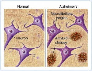
Alzheimer’s memory problems appear to be caused by the formation of two major tissues in the brain – plaques and tangles. Plaques form when beta-amyloid proteins clump together, blocking cell-to-cell signaling. According to Cummings, plaque appears to come first in the degeneration process, but these clusters do not seem to damage the brain’s cells. Tangles are twisted strands of the protein tau. When they begin to form, nerve cells begin to die, causing memory failure, personality changes and other symptoms of the disease.

Currently, most drugs have been aimed at preventing plaque buildup, but the high failure rate of these drugs suggests the need for a better understanding of the disease itself, as well as a broader focus on other drug targets, such as the cell-killing tangles.

“I would argue that just as you would diversify an investment portfolio, you need to diversify [the drug development] portfolio to ensure there’s a chance for success,” Cummings said.

Another consideration is the economic impact of Alzheimer’s disease. According to the Alzheimer’s Association, in 2014, the estimated cost to Americans for the care of those of Alzheimer’s will be $214 billion— and an estimated $1.2 trillion in 2050.

“We are investing about $600 million per year in Alzheimer’s research and about $6 billion per year in cancer research… at the same time that Alzheimer’s is having a larger impact on the U.S. economy,” Cummings said. “That doesn’t mean we should be doing less cancer research; we should be doing more Alzheimer’s research.”

Lack of standardization among clinical trials is another roadblock to drug testing success, as inconsistent procedures can make data appear to be more or less successful, Cummings noted.

So far, five drugs have been approved for the treatment of Alzheimer’s, yet they only treat symptoms of the disease, not the underlying plaque and tangle process. One explanation for this is that scientists better understand the mechanics of Alzheimer’s symptoms, making it technically more difficult to develop drugs that modify the disease.

“Our new science is pointing us toward the plaque and tangle process; if we could arrest that process particularly early, we could keep people at a very high functional level,” Cummings said.

Additionally, Cummings pointed out that it takes 10 years for a drug to make it through the pipeline— meaning the science of drugs being tested now is already a decade behind. However, the protocol for testing procedures is unchangeable, meaning this delay is an inherent part of the drug development problem.

Developing drugs to combat Alzheimer’s disease is a multi-faceted and complex process, and these researchers hope their findings will highlight the importance of increased efforts.

“Overall, my message is we’re doing too little, investing too little; we need the help of the government, philanthropists, advocacy groups, venture capital,” Cummings said. “We need a very comprehensive approach to developing new treatments for Alzheimer’s disease, because it’s truly in a disastrous state.”

Source: fox news


Working in the Night Shift Disrupts Brain Clock

Working the Night Shift Disrupts Brain Clock

It is already known that working the graveyard shift can negatively impact health, and the body of evidence continues to grow. Now, researchers have shown that forced nocturnalism may affect the brains’ internal clock in a way that’s similar to Alzheimer’s disease.

The circadian system in mammals orchestrates the daily rhythms of activity and sleep with day and night. Disrupting this cycle throws off physiological balance, and can lead to increased risks of heart disease and diabetes in people.

But until now little has been known about how shift work alters the brain’s master timekeeper.

Simulating the Graveyard Shift

To test the effects of working late, researchers put rats through a simulated work week. Seven rats worked during their typical nocturnal active period, while 12 others were made to work days instead. Both sets of rats logged a typical 5-day work week with weekends, and stayed on the grind for five weeks.

Researchers simulated “work” by placing rats on slowly rotating drums, which required the rats to stay awake and move, but didn’t overexert them. After their shifts, researchers monitored rats’ movements with motion sensors to test for abnormalities.

Within two weeks, rats working the graveyard shift gained a significant amount of weight as their post-work activity decreased. Their activity levels were also more erratic, fluctuating from unpredictable highs to lows. Rats that worked a typical week didn’t change their behavior. Further, a two-day weekend wasn’t enough time to recover from behavioral changes resulting from working the night-shift.

Researchers determined by measuring post-work activity levels that these changes occurred in rats’ biological clock, or master circadian pacemaker, which is located within the brain. The disruption was so pronounced that the rats’ behavior resembled animals that had had that region severed. For a human analogy, the rats’ symptoms stemming from a dysfunctional circadian clock were similar to a person diagnosed with dementia.

Researchers published their findings Tuesday in the Journal of the Royal Society Interface.

Source: discover


‘Chaperone’ compounds could help treat Alzheimer’s

health-genetics-alzheimers-369588Researchers have devised a wholly new approach to the treatment of Alzheimer’s disease involving the so-called retromer protein complex.

Retromer plays a vital role in neurons, steering amyloid precursor protein (APP) away from a region of the cell where APP is cleaved, creating the potentially toxic byproduct amyloid-beta, which is thought to contribute to the development of Alzheimer’s.

Using computer-based virtual screening, the researchers identified a new class of compounds, called pharmacologic chaperones that can significantly increase retromer levels and decrease amyloid-beta levels in cultured hippocampal neurons, without apparent cell toxicity.

Dagmar Ringe, PhD, Harold and Bernice Davis Professor in the Departments of Biochemistry and Chemistry, said their challenge was to find small molecules-or pharmacologic chaperones-that could bind to retromer’s weak point and stabilize the whole protein complex.

This was accomplished through computerized virtual, or in silico, screening of known chemical compounds, simulating how the compounds might dock with the retromer protein complex. (In conventional screening, compounds are physically tested to see whether they interact with the intended target, a costlier and lengthier process.)

The screening identified 100 potential retromer-stabilizing candidates, 24 of which showed particular promise. Of those, one compound, called R55, was found to significantly increase the stability of retromer when the complex was subjected to heat stress.

The researchers then looked at how R55 affected neurons of the hippocampus, a key brain structure involved in learning and memory.

More important, a subsequent experiment showed that the compound significantly increased retromer levels and decreased amyloid-beta levels in cultured neurons taken from healthy mice and from a mouse model of Alzheimer’s. The researchers are currently testing the clinical effects of R55 in the actual mouse model.

The study has been published online in the journal Nature Chemical Biology.

Source: zee news


IMS-BHU researchers identify novel drug target for treating Alzheimer’s disease

Researchers at the Institute of Medical Sciences, Banaras Hindu University (IMS-BHU) have identified RhoA as a novel drug target in the treatment of Alzheimer’s disease. The study has been published in the FASEB Journal.

Dr Debabrata Dash, professor and head, department of biochemistry, IMS-BHU, said, “We have discovered a novel drug target of amyloid beta toxicity and Alzheimer’s disease in platelet model. We have found that amyloid beta exercises its activity on a cell through activation of a small GTP-binding protein known as RhoA. When we inhibit RhoA activity by employing pharmacological inhibitors, the toxic effects of amyloid beta are prevented. This information would have significant implications in drug development against Alzheimer’s disease.”

Alzheimer’s disease is the most common cause of cognitive decline and memory loss in the elderly. Although local deposit of a small peptide called ‘amyloid-beta’ is known to be responsible for Alzheimer pathology, the underlying molecular mechanism remains largely obscure. This is the reason why there is no effective therapy against this highly debilitating condition.

Platelets are blood cells that are responsible for stoppage of bleeding at the site of injury. Interestingly, platelets are a major source of amyloid-precursor protein in blood.

Dr Dash and co-workers (Vijay Sonkar and Paresh Kulkarni) at Banaras Hindu University have identified target molecules of amyloid-beta in cells using platelets as peripheral model of neurons.

Their study showed that amyloid-beta was able to strongly stimulate platelets leading to aggregate formation. Intravenous administration of the peptide in mouse accelerated thrombus formation in pulmonary vessels.

The effect of amyloid-beta on platelets was found to be mediated through activation of RhoA, a small GTP-binding protein responsible for cytoskeletal reorganization in cells, and that inhibition of RhoA by a specific pharmacological agent reversed the effects of amyloid-beta on platelets.

In order to understand molecular underpinnings amyloid action, researchers went further to demonstrate phosphorylation of downstream effectors of RhoA, namely MYPT1 and myosin light chain, when the cells were exposed to amyloid-beta.

According to the researchers, patients of Alzheimer’s also have clotting abnormalities, which could be explained by amyloid-beta-induced activation of platelets.

“The findings of this study thus identify RhoA as a novel drug target in the treatment of Alzheimer’s disease, and unravel the possible cause of clotting abnormalities seen in these patients,” said Dr Dash.

Sourrce: India Medical Times


New drug target for Alzheimer’s identified

Researchers have identified abnormal expression of genes, resulting from DNA relaxation, that can be detected in the brain and blood of Alzheimer’s patients.
The protein tau is involved in a number of neurodegenerative disorders, including Alzheimer’s disease.

Previous studies have implicated DNA damage as a cause of neuron, or cell, death in Alzheimer’s patients.

Given that DNA damage can change the structure of DNA within cells, the researchers led by Bess Frost examined changes in DNA structure in tau-induced neurodegeneration.

They used transgenic flies and mice expressing human tau to show that DNA is more relaxed in tauopathy.

They then identified that the relaxation of tightly wound DNA and resulting abnormal gene expression are central events that cause neurons to die in Alzheimer’s disease.

“Our work suggests that drugs that modify DNA structure may be beneficial for treating Alzheimer’s Disease,” they wrote.

The study is published in the journal Nature Neuroscience.

Source: Business Standard


Cholesterol linked to Alzheimer’s protein, unclear why

Patterns of “good” and “bad” cholesterol usually associated with heart risks also predicted the levels of Alzheimer’s-related beta amyloid protein seen in the brains of study participants.

“One of the important themes emerging from dementia research over the past 15 years is that there are intriguing connections between vascular disease and Alzheimer’s disease,” Bruce Reed, who led the research, told Reuters Health by email.

Reed is a professor and associate director of the University of California Davis Alzheimer’s Disease Research Center.

“It has become increasingly clear that what have been traditionally thought of as vascular risk factors – things like hypertension, diabetes and elevated cholesterol – are also risk factors for Alzheimer’s disease,” Reed said.

In previous work, Reed and his colleagues found a connection between overall vascular risk and levels of brain amyloid.

“Amyloid deposition is important because it is widely believed by scientists to be a key event that initiates a chain of events that eventually, years later, results in the dementia of Alzheimer’s disease,” Reed said.

“There was also previous work in cell cultures and with animals that suggested that cholesterol plays an important role in promoting the deposition of amyloid in the brain,” Reed said.

For the new study, published in JAMA Neurology, the researchers examined the cholesterol levels of 74 elderly people who had normal to mildly impaired cognitive function. Researchers also measured brain deposits of beta amyloid protein with positron emission tomography (PET scanning).

They found that on average, participants who had higher levels of the “bad” LDL cholesterol and lower levels of “good” HDL cholesterol also had higher levels of amyloid in the brain.

Elevated LDL-cholesterol is associated with cardiovascular disease while high levels of HDL-cholesterol are thought to protect against heart disease.

“We think this is a very important finding, but as with all novel findings it needs to be replicated,” Reed said. “Assuming that the basic pattern is found in other groups of patients, it is urgent that we try to understand the mechanism(s) behind this finding.”

“Cholesterol in blood and cholesterol in brain are separate ‘pools,’ walled off from one another by the blood brain barrier. We measured cholesterol in blood. So that is one question that needs to be answered – how do cholesterol levels in blood and in brain influence each other,” Reed said.

“And in the brain, it is not entirely understood how changing cholesterol levels might reduce amyloid deposition. We are very interested in the idea that higher HDL (‘good’) cholesterol levels may help the brain more efficiently clear the toxic amyloid at an early stage,” Reed said.

“If those questions were understood we could begin to think about how to change cholesterol levels so as to prevent the buildup of amyloid,” Reed added.

In other studies, some researchers have found evidence that LDL cholesterol-lowering drugs, such as statins, might offer some protection against Alzheimer’s disease, but the results have been inconsistent.

Reed’s team didn’t see any associations between current use of cholesterol medication by the participants and their amyloid levels.

In their report, Reed and his coauthors caution that the study does not prove cholesterol is directly affecting amyloid deposition. For instance, they write, unhealthy cholesterol could be linked to vascular damage, such as small strokes, and those micro-injuries could be the reason for the protein deposits.

He encourages people to follow the advice of their doctor and try to achieve the cholesterol guidelines set by the American Heart Association.

“A remarkable number of people who are alive now will live into their 80s or beyond – the period of highest risk for Alzheimer’s. This study is one more piece of evidence that what we do now can shape our health positively in those years,” Reed said.

Source: ABS CBN news


Vitamin E may slow spread of mild-to-moderate Alzheimer’s disease

Researchers say vitamin E might slow the progression of mild-to-moderate Alzheimer’s disease — the first time any treatment has been shown to alter the course of dementia at that stage.

In a study of more than 600 older veterans, high doses of the vitamin delayed the decline in daily living skills, such as making meals, getting dressed and holding a conversation, by about six months over a two-year period.

The benefit was equivalent to keeping one major skill that otherwise would have been lost, such as being able to bathe without help. For some people, that could mean living independently rather than needing a nursing home.

Vitamin E did not preserve thinking abilities, though, and it did no good for patients who took it with another Alzheimer’s medication. But those taking vitamin E alone required less help from caregivers — about two fewer hours each day than some others in the study.

“It’s not a miracle or, obviously, a cure,” said study leader Dr. Maurice Dysken of the Minneapolis VA Health Care System. “The best we can do at this point is slow down the rate of progression.”

The U.S. Department of Veterans Affairs sponsored the study, published Tuesday by the Journal of the American Medical Association.

No one should rush out and buy vitamin E, several doctors warned. It failed to prevent healthy people from developing dementia or to help those with mild impairment (“pre-Alzheimer’s”) in other studies, and one suggested it might even be harmful.

Still, many experts cheered the new results after so many recent flops of once-promising drugs.

“This is truly a breakthrough paper and constitutes what we have been working toward for nearly three decades: the first truly disease-modifying intervention for Alzheimer’s,” said Dr. Sam Gandy of Mount Sinai School of Medicine in New York. “I am very enthusiastic about the results.”

About 35 million people worldwide have dementia, and Alzheimer’s is the most common type. In the U.S., about 5 million have Alzheimer’s. There is no cure and current medicines just temporarily ease symptoms.

Researchers don’t know how vitamin E might help, but it is an antioxidant, like those found in red wine, grapes and some teas. Antioxidants help protect cells from damage that can contribute to other diseases, says the federal Office on Dietary Supplements. Many foods contain vitamin E, such as nuts, seeds, grains, leafy greens and vegetable oils. There are many forms, and the study tested a synthetic version of one — alpha-tocopherol — at a pharmaceutical grade and strength, 2,000 international units a day.

Years ago, another study found that the same form and dose helped people with more advanced Alzheimer’s, and many were prescribed it. But vitamin E fell out of favor after a 2005 analysis of many studies found that those taking more than 400 units a day were more likely to die of any cause.

The new study involved 613 veterans, nearly all male, 79 years old on average, with mild to moderate Alzheimer’s, at 14 VA centers. All were already taking Aricept, Razadyne or Exelon — widely used, similar dementia medicines.

Participants were placed in four groups and given either vitamin E, another dementia medicine called memantine (its brand name is Namenda), both pills or dummy pills.

After a little more than two years of follow-up, those on vitamin E alone had a 19 percent lower annual rate of decline in daily living skills compared to the placebo group. Memantine made no difference, and vitamin E did not affect several tests of thinking skills.

“It’s a subtle effect but it’s probably real,” Dr. Ron Petersen, the Mayo Clinic’s Alzheimer’s research chief, said of the benefit on daily living from vitamin E. “That has to be weighed against the potential risks” seen in earlier studies, he said.

Heather Snyder, director of medical and scientific operations for the Alzheimer’s Association, said the group’s position is that “no one should take vitamin E for Alzheimer’s disease or other memory issues except under the supervision of a physician,” because it can interfere with blood thinners, cholesterol drugs and other medicines.

The new results also need to be verified in a fresh study that includes more women and minorities, she said.

Source: fox news


Study: Concussions May Lead To Alzheimer’s Plaque Buildup For Some

Concussions have already been linked to the Alzheimer’s-like degenerative brain disease chronic traumatic encephalopathy (CTE) in athletes and military members who have experienced repeated head blows and traumatic brain injuries.

Now, a new study links concussions to Alzheimer’s disease itself.

Mayo Clinic researchers gave brain scans to 141 Minnesotans who had been experiencing memory problems, and found those who had suffered a brain injury that caused them to black-out had more amyloid plaques in their brain than those who hadn’t.

Amyloid plaque is the telltale sign of Alzheimer’s disease, formed by pieces of a sticky protein that break off in the brain and clump together. Some clumps may form in brain regions involved in learning, memory and thinking, the Alzheimer’s Association explains. More plaques form as the disease progresses.

Researchers gave brain scans to 448 people without any memory or cognitive problems, and 141 people who had mild cognitive impairment (MCI), a condition characterized by declines in memory and thinking skills that aren’t caused by aging. They were also asked whether they had ever experienced a brain injury that caused them to lose consciousness. People with MCI are at a heightened risk of developing Alzheimer’s or another type of dementia, but not everyone with the condition will get worse.

The researchers found 18 percent of those with MCI had reported a prior brain injury, and on scans, they saw the patients had an average of 18 percent more amyloid plaques than those with no history of head trauma. They found no plaque differences in any of the brain scans of people without memory problems, regardless of whether they’d had a brain injury.

Source: dig triad