Celery cure for leukemia

Celery cure for leukemia

Eating foods like celery and parsley which contain natural flavonoid apigenin, may prevent leukemia, concluded by Dutch scientists from the University of Groningen .

According to Maikela Pepelenbosh, one of the participants in the research, tests showed that apigenin – otherwise common ingredient in fruit and vegetables – is able to terminate the development of two types of leukemia cells and reduce their chances of survival .

On the other hand, the team found that apigenin may reduce the positive effects of chemotherapy treatment at people with leukemia. Flavonoids are compounds with antioxidant properties that protect cells from damage caused by the oxygen molecules .
It is important to emphasize that the celery is rich in vitamins A, B, C and E, and heals and helps with diseases caused by a lack of these vitamins,

  • -gout and rheumatism,
  • -stimulate the secretion of urine,
  • -throws sand and stones in the kidneys and bladder,
  • -leaflets bronchitis attacks fear and spasms in the chest,
  • -various skin diseases,
  • -acts against temperature,
  • -stimulates circulation,
  • -affecting the growth and care of the hair and the most powerful aphrodisiac.

WARNING: Patients suffering from kidney inflammation should not take celery, and is not recommended for pregnant women . Heart patients should not use celery in small quantities, as a common seasoning.

Celery cure for leukemi2a

Improves blood count also blood circulation, improve appetite, and eliminates interference with digestion. Acetylenic is one of its ingredients and it help to stops the reproduction of cancer cells.

Celery contain substances that act on relaxing the artery, allowing them to proliferate, reducing the amount of stress hormones, which also influences the contraction of blood vessels.

Celery reduces bad cholesterol, because of the abundance of Vitamin C helps to reduce the symptoms of fly and improve immune response. This is because of its composition containing phthalide of the active ingredients which relax the muscles of the arteries and thus regulate blood pressure.

Celery cure for leukemi3

Cholesterol lowering effect is achieved best with celery juice, but it must be persistent because they are expected effect may be seen only after eight weeks.

Some previous studies have shown that apigenin, which has in celery, parsley, red wine, tomato juice and various other herbs may be beneficial in the prevention of ovarian cancer .

Source: secretly healthy


Biochemical cascade causes bone marrow inflammation and blood disorders

Biochemical cascade causes bone marrow inflammation and serious blood disorders

Like a line of falling dominos, a cascade of molecular events in the bone marrow produces high levels of inflammation that disrupt normal blood formation and lead to potentially deadly disorders including leukemia, an Indiana University-led research team has reported.

The discovery, published by the journal Cell Stem Cell, points the way to potential new strategies to treat the blood disorders and further illuminates the relationship between inflammation and cancer, said lead investigator Nadia Carlesso, M.D., Ph.D., associate professor of pediatrics at the Indiana University School of Medicine.

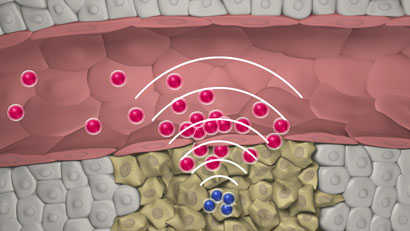
Bone marrow includes the cells that produce the body’s red and white blood system cells in a process called hematopoiesis. The marrow also provides a support system and “home” for the blood-producing cells called the hematopoietic micro-environment. The new research demonstrates the importance of the hematopoietic micro-environment in the development of a group of potentially deadly diseases called myeloproliferative disorders.

“It has been known for years that there are links between inflammation and cancer, but these studies have been challenged by the lack of genetic models, especially for blood-based malignancies,” said Dr. Carlesso, a member of the hematologic malignancy and stem cell biology program within the Wells Center for Pediatric Research at IU.

The researchers focused on what happens when there are abnormally low levels of a molecule called Notch, which plays an important role in the process of blood cell production. Using a genetically modified mouse, they found that the loss of Notch function in the microenvironment causes a chain of molecular events that result in excess production of inflammatory factors.

The high levels of inflammation in the bone marrow were associated with the development of a myeloproliferative disorder in the mice. Myeloproliferative diseases in humans can result in several illnesses caused by overproduction of myeloid cells, which are normally are used to fight infections. These diseases can put patients at risk for heart attack or stroke, and frequently progress into acute leukemia and bone marrow failure, which have fatal outcomes. Unfortunately, there are no effective therapies for the majority of myeloproliferative diseases.

When Dr. Carlesso’s team blocked the activity of one of the molecules in this biochemical cascade, the myeloproliferative disorder in the mice was reversed. In addition, elevated levels of the blocked molecule were found in samples from human patients with myeloproliferative disease. These findings suggest that developing drugs that target this inflammatory reaction at different key points could be a promising strategy to limit the development of myeloproliferative disease in humans.

The molecular cascade leading to inflammation was not occurring directly in the bone marrow cells that produce blood cells, but in cells of the bone marrow microenvironment, especially in endothelial cells that line the capillaries — tiny blood vessels — inside the bone marrow. This was a key discovery, Dr. Carlesso said.

“This work indicates that we need to target not only the tumor cells, but also the inflammatory microenvironment that surrounds them and may contribute to their generation,” she said.

“We believe that this combined strategy will be more effective in preventing myeloproliferative disease progression and transformation in acute leukemias.” Dr. Carlesso also noted that the Notch molecule is mostly known as an oncogene — one that can cause cancer — and so is often targeted by therapies for other types of cancer. The new research indicates that clinicians need to be aware of the effects that reducing levels of Notch function could have on the blood development process, she said.

Source: science daily


Drowning Prevention: How to Be Cautious Around Water

Drowning Prevention How to Be Cautious Around Water

Whether you are taking your kids to a pool, a river or the ocean, it is crucial that you exercise caution and use good judgment about water safety. Drowning is a very important cause of injury-related death among young children. Although young children are particularly at risk, even older kids who know how to swim can experience difficulty, or even drown.

When you’re out with your kids having fun in the water, safety issues may not be at the top of your mind. However, in order to keep your kids safe, you need to follow some basic precautions. By adopting proper water safety practices, you can help protect your family from serious, even deadly, accidents and injuries.

Ensure Adult Supervision
Though you may feel your kids are old enough to monitor their own activities in the water, don’t let your guard down. Whenever children are near water, responsible adults should closely supervise them at all times.

When watching children – whether near or around a pool, river, ocean, or even a bathtub – avoid multitasking. Do not engage in distracting activities that can cause you to lose your focus on watching your children, such as making phone calls, reading, playing games with other adults, or watching TV.

Wear Life Jackets
Life jackets aren’t just for boating. When your kids are around natural bodies of water – such as rivers or the ocean – consider if life jackets should be mandatory. Even if your kids are excellent swimmers, it’s possible for rip tides and currents to overpower them. Children who are weaker swimmers may need to wear a life jacket when they are in or near swimming pools as well.

Use Pool Fences
Swimming pools, however small, inside houses can be as dangerous as the ocean when it comes to small children. Even if parents are nearby, children are at risk for drowning if proper precautions aren’t taken.

If you have a swimming pool in your house, it is recommended to separate your house from the pool by installing an “isolation fence.” This fence should be four-sided to completely encompass the pool, and should include gates that self-close and self-latch. Isolation fences can help keep children away from the pool area when they’re in the surroundings playing.

Know the Basics
Sometime’s prevention isn’t enough, and accidents happen. It is recommended that all parents learn basic life-saving skills. In addition to knowing how to swim (or at least how to float and move through the water), adults with young children should also learn how to administer cardiopulmonary resuscitation (CPR). It isn’t hard to find a basic CPR course. There are many hospitals and NGOs offering the basic CPR courses to interested individuals.

CPR helps to save lives by keeping a flow of oxygenated blood to vital organs. When the heart stops, a person can die in a matter of minutes. If you’re trained in CPR, and administer your skills promptly, you may be able to help save a life.

Avoid Disaster: Use Caution
According to an estimate, India has the highest number of child deaths due to drowning. One of the most tragic aspects of these deaths is that they are completely avoidable. Water injuries and deaths can be prevented, as long as adults remain mindful of children at play. It’s important not to become careless around water, even when your kids are splashing and having fun. By learning the best practices for water safety at pools, rivers, and ocean beaches, you can help keep your kids safe and healthy.

Source: healthline


Honeydew melons play role in boosting brain health and more

Honeydew melons play role in boosting brain health and more

Honeydew melon is more than a mildly sweet, refreshing melon typically enjoyed during the warmer months; it’s also a highly nutritious food that can help maintain a healthy weight, fight colds, keep blood pressure in check and keep nervous system health functioning well.

Here’s a closer look at the top health benefits of honeydew melon. As always, it’s wise to choose organic foods as they’re free of harmful pesticides that fill the body with toxins.

Health benefits of honeydew melon

1) Weight control. Honeydew melons have only 60 calories for every 1/2 cup making it a low-calorie option that also satisfies the craving for something sweet. Furthermore, they have a high water content that not only helps with hydration, but provides a feeling of fullness to stave off the urge to eat foods that may be unhealthy.

2) Fights colds. These tasty melons provide approximately half of daily vitamin C needs in just one cup. Since vitamin C plays a role in keeping the immune system strong, honeydew can help fight infection and ward off illnesses.

3) Regulates blood pressure. Nutrition experts at the University of Texas Southwestern (UTS) Medical Center at Dallas advise eating melons like honeydew, citing their high potassium levels as a way to reduce salt retention and therefore, better control blood pressure. It’s also thought that the ratio of water to potassium on honeydew can help lower blood pressure may also help prevent an increase in blood pressure.

4) Healthy nervous system. Honeydew contains B vitamins thiamin and niacin which are known to not only increase energy levels, but to remove toxins from the body and in turn, boost nervous system health. The vitamins have been known to help boost brain health and prevent detrimental changes to it such as the onset of Alzheimer’s.

Honeydew may be eaten fresh, directly scooped from the melon itself as a snack, or can be added to a variety of dishes including fruit or green salads.

Source: Natural news


Working in the Night Shift Disrupts Brain Clock

Working the Night Shift Disrupts Brain Clock

It is already known that working the graveyard shift can negatively impact health, and the body of evidence continues to grow. Now, researchers have shown that forced nocturnalism may affect the brains’ internal clock in a way that’s similar to Alzheimer’s disease.

The circadian system in mammals orchestrates the daily rhythms of activity and sleep with day and night. Disrupting this cycle throws off physiological balance, and can lead to increased risks of heart disease and diabetes in people.

But until now little has been known about how shift work alters the brain’s master timekeeper.

Simulating the Graveyard Shift

To test the effects of working late, researchers put rats through a simulated work week. Seven rats worked during their typical nocturnal active period, while 12 others were made to work days instead. Both sets of rats logged a typical 5-day work week with weekends, and stayed on the grind for five weeks.

Researchers simulated “work” by placing rats on slowly rotating drums, which required the rats to stay awake and move, but didn’t overexert them. After their shifts, researchers monitored rats’ movements with motion sensors to test for abnormalities.

Within two weeks, rats working the graveyard shift gained a significant amount of weight as their post-work activity decreased. Their activity levels were also more erratic, fluctuating from unpredictable highs to lows. Rats that worked a typical week didn’t change their behavior. Further, a two-day weekend wasn’t enough time to recover from behavioral changes resulting from working the night-shift.

Researchers determined by measuring post-work activity levels that these changes occurred in rats’ biological clock, or master circadian pacemaker, which is located within the brain. The disruption was so pronounced that the rats’ behavior resembled animals that had had that region severed. For a human analogy, the rats’ symptoms stemming from a dysfunctional circadian clock were similar to a person diagnosed with dementia.

Researchers published their findings Tuesday in the Journal of the Royal Society Interface.

Source: discover


5 Tricks For Easy, Healthy Breakfasts

5 Tricks For Easy, Healthy Breakfasts

You may have seen reports in the news lately questioning the benefits of breakfast for weight loss, but I’m not ready to sanction skipping. In my experience, eating breakfast strongly supports weight control, and several studies back what I’ve seen in my 15+ years of counseling clients – breakfast fuels your body when you’re most active, and therefore most likely to burn off what you’ve eaten. It also tends to prevent late night overeating, when you’re less active, and more prone to racking up a fuel surplus that feeds fat cells.

Also, weight loss aside, “breaking the fast” is a savvy nutrition strategy, because it’s a chance to fit in servings of produce, whole grains, healthy fats, and lean proteins. Missing that opportunity, particularly day after day, can lead to shortfalls that deprive your body of important health protective nutrients.

Eating breakfast, especially one with protein, is also a smart way to build and maintain metabolism-boosting muscle. One recent study, published in the Journal of Nutrition, found that muscle building was 25% greater among people who ate a diet with an evenly distributed protein intake, compared to those who consumed less protein at breakfast, slightly more at lunch, and the majority of their protein at dinner.

Finally, a study published last year from the American Heart Association found that over a 16 year period, regular breakfast skippers had a 27% higher risk of a heart attack or fatal heart disease.

If you’re on board for a daily breakfast, but your biggest barrier is time, here are five tips and tricks to help you create shortcuts, so you can reap the benefits without running late.

Chill your oatmeal
Oatmeal doesn’t have to be served warm. Cook, then chill individual portions, and stash them in the fridge in small containers you can grab, along with a spoon, on your way out the door. Just mix a protein powder (like pea, hemp, or organic whey) into rolled oats, add hot water, stir, fold in fresh fruit, cinnamon, and nuts, and chill. Or skip the protein powder, and mix the oats, fruit, cinnamon, and nuts into nonfat organic Greek yogurt, and chill to make a grab-n-go mueslix.

Hard boil it
Many of my clients enjoy omelets on the weekends, but feel like an egg-based breakfast takes too much time during the week. For a make-ahead option, prep hard boiled organic eggs on a Sunday for the upcoming week. While you’re making dinner, take a few extra minutes to whip up a simple egg salad for breakfast the next morning. Mix chopped egg with either guacamole or pesto, diced or shredded veggies, and a small scoop of cooked, chilled quinoa or brown rice. Grab a portion with a fork in the a.m., and you’re good to go.

Have dinner for breakfast
It may seem odd to chow down on a garden salad topped with lentils or salmon at 8 am, but who says breakfast meals have to look different than lunch or dinner? Many of my clients make double portions in the evening, and eat seconds for breakfast the next day. Give it a try – you may just find that warmed up stir fry, veggie “pasta” or a crisp entrée salad is your new favorite way to start the day.

Pre-whip your smoothie
Smoothies are pretty fast, but I know that when you’re running late, just tossing ingredients into a blender and pressing a button can require more time than you can spare. If that tends to be the case, blend up a smoothie just before bed, stash it in a sealed to-go jug in the fridge, grab it on your way out the door, and shake it up before sipping. If you’re a chocolate lover, but you want to sneak some greens into your first meal of the day, check out my chocolate cherry kale smoothie recipe.

Make a meal out of snack foods
It’s perfectly OK to cobble together a breakfast from an assortment of snack foods, including veggies with hummus and whole grain crackers, or trail mix made from nuts or seeds, unsweetened preservative-free dried fruit, and a whole grain cereal you can eat with your hands. For more easy and energizing ideas, check out Health’s snack recipe database. Bon (breakfast) appetit!

Source: health


Top 10 Food-Borne Parasites of Greatest Global Concern : Africa

Top 10 Food-Borne Parasites of Greatest Global Concern

A new report released today by two United Nations agencies identifies a “top 10” of food-borne parasites with the greatest global impact, including those found in pork, fish, fresh produce, fruit juice and milk, among other foods.

According to the report, produced by the Food and Agriculture Organization (FAO) and the World Health Organization (WHO), the top ten are:

  • Taenia solium (pork tapeworm): In pork
  • Echinococcus granulosus (hydatid worm or dog tapeworm): In fresh produce
  • Echinococcus multilocularis (a type of tapeworm): In fresh produce
  • Toxoplasma gondii (protozoa): In meat from small ruminants, pork, beef, game meat (red meat and organs)
  • Cryptosporidium spp. (protozoa): In fresh produce, fruit juice, milk
  • Entamoeba histolytica (protozoa): In fresh produce
  • Trichinella spiralis (pork worm): In pork
  • Opisthorchiidae (family of flatworms): In freshwater fish
  • Ascaris spp. (small intestinal roundworms): In fresh produce
  • Trypanosoma cruzi (protozoa): In fruit juices

“Obviously this top ten is a more general, global perspective and does not necessarily reflect parasite rankings at a national level where each country may have more precise information,” Renata Clarke, head of food safety and quality at FAO, said in a news release.

“But considering the problems they cause, these parasites do not get the attention they deserve. We hope that by releasing a top ten ranking we can increase awareness among policy makers, the media and the general public about this major public health issue,” she added.

The parasites affect the health of millions of people every year, infecting muscle tissues and organs, causing epilepsy, anaphylactic shock, amoebic dysentery and other problems, the agencies noted. Some can live on in people’s bodies for decades.

Despite their huge social costs and global impacts, information is generally lacking regarding just where these parasites come from, how they live in the human body, and – most importantly – how they make people sick.

The list and report were developed following a request by the global food standards body, the Codex Alimentarius Commission (Codex), for FAO and WHO to review the current status of knowledge on parasites in food and their public health and trade impacts.

The Codex Committee on Food Hygiene is now developing new guidelines for the control of these parasites. FAO and WHO are supporting the process by providing scientific and technical information.

The aim is to develop new standards for the global food trade that will help countries control the presence of these parasites in the food chain.

The report lists a number of ways to reduce the risk of parasite infections. For farmers, it advises the use of organic fertilizer, particularly on produce, should be closely monitored to ensure it is composted properly and all faecal matter is removed. Water quality must also be closely monitored.

For consumers, it advises that all meat should be well cooked and only clean water should be used to wash and prepare vegetables.

The agencies noted that in Europe, more than 2,500 people are affected by food-borne parasitic infections each year. In 2011, there were 268 cases of trichinellosis and 781 cases of echinococcosis recorded in the European Union.

In Asia, there is no precise national data but parasitic diseases are known to be widely spread and are recognized as major public health problems in many countries.

Meanwhile, there is no data at all in most African nations on the prevalence of food-borne parasites in humans because of a general lack of surveillance systems.

In the United States, Neurocysticercosis, caused by Taenia solium, is the single most common infectious cause of seizures in some areas of the US where 2,000 people are diagnosed with neurocysticercosis every year. Toxoplasmosis is a leading cause of food-borne illness and death.

Source: All Africa


‘Oxygen therapy is like a prescription drug, should be used rationally’

‘Oxygen therapy is like a prescription drug, should be used rationally’

An optimum amount of oxygen is essential for the functioning and survival of all body tissues and even a few minutes deprivation can prove fatal. When saturation level of oxygen in the body falls due to some respiratory illness or injury then we need to replenish it artificially to maintain an optimum level by giving oxygen therapy to the patient. This method of dealing with ‘respiratory failure’ was explained succinctly by Dr Girija Nair, head of the pulmonary medicine department at Dr D Y Patil Medical College, Mumbai on the 1st day of 20th NESCON (National Conference on Environmental Sciences and Pulmonary Diseases), a 3-day event which started on Friday and is being organized here by the Academy of Respiratory Medicine under the auspices of Environmental Medical Association.

Oxygen therapy is one of the key treatments in respiratory care. This therapy is used to treat documented hypoxemia (as in case of smokers), severe respiratory distress, severe trauma, interstitial lung disease (ILD), chronic obstructive pulmonary disease (COPD) including chronic asthma and chronic bronchitis, pulmonary hypertension, acute myocardial infarction, and chronic lung patients during exercise. Some may need it for long term oxygen therapy at home (LTOT).

Before prescribing oxygen therapy there should be a clear indication of its need and the type of therapy needed. The doctor should list the purpose of the therapy, demonstrate the procedure and also list possible complications. For safe and effective treatment with this therapy, it should be remembered that the oxygen used in oxygen therapy is a prescription drug which is to be used under a doctor’s prescription and not like the oxygen we breathe naturally. The prescription should clearly state the airflow rate, delivery system, duration and instructions for monitoring. Regular evaluation of patient is necessary.

The basic purpose to ventilate patients through oxygen therapy is to improve gas exchange till lung functions improve. Oxygen saturation has to be within normal limits. Target should be PaO2 (pressure) not less than 60mm Hg and SpO2 (saturation pressure) not less than 90mm Hg. Excess supply can do more harm than good and one should ensure that this does not happen. Hundred per cent Fi02 breathing is associated with decreased ventilation (obstruction).

Dr Nair shared her experience of having a tough time convincing the ICU staff to lower the oxygen flow while administering to patients of COPD requiring low flow oxygen, as COPD patients may worsen with high flow oxygen. Patients with chronic lung disease should never be delivered at more than 2-3 litre/minute oxygen.

Oxygen needed in the therapy can be dispensed/supplied from: piped in oxygen cylinders which can supply oxygen for up to 57 hours at the desired regulated flow; oxygen concentrator which has molecular sieves to supply 90% oxygen, and permeable membrane to supply 40% oxygen; and liquid oxygen which can be used for 7 days at 2 litres/minute; can be refilled, weighs less, but is costly and can cause thermal burns as liquid oxygen has a very low temperature.

Any system of oxygen delivery would require the following — oxygen supply, flowmeter, oxygen tubing, delivery device and a humidifier (if need be). There are (1) low flow systems which do not provide constant flow oxygen and contribute partially to inspired gas the patient breathes. Examples are nasal cannula, simple mask, non/partial re-breather masks and (2) high flow systems which deliver specific and constant percentage of oxygen independent of patient’s breathing. Examples are venturi mask, tracheostomy collar mask, and T piece.

Venturi mask mixes a specific volume of air and oxygen and is used for accurate delivery of low concentrations of oxygen between 24-60% at a flow of 4-15 litre/minute. The mask is so constructed that there is constant flow of room air blended with a fixed concentration of oxygen. Valves are colour coded and flow rate required to deliver a fixed concentration is shown on each valve. Each colour code corresponds to a precise oxygen concentration and a specific flow rate.

Nasal cannula is a disposable plastic device (having 2 protruding prongs) and connected to an oxygen source for delivering low-medium concentrations of oxygen — 24-44% at flow rate of 1 litre/minute. They are easy and comfortable to use, but some times prolonged use may cause nasal irritation and pharyngeal mucosa.

The simple face mask is made of clear flexible plastic or rubber that can be moulded. It delivers 35-60% oxygen at a flow rate of 6-10 litre/minute. It is used when an increased delivery of oxygen is needed for short duration of time less than 12 hours. It is not very patient-friendly and requires frequent monitoring to check if it is placed correctly.

Partial re-breather mask has an oxygen reservoir bag which should remain inflated during inspiration and expiration. It can deliver oxygen concentrations up to 80%. The oxygen flow rate is maintained at a minimum of 6 litre/minute to ensure that the patient does not re-breathe large amounts of exhaled air. The remaining exhaled air exits through vents.

The non re-breather mask also has an oxygen reservoir bag. It provides highest possible concentration of oxygen of 95-100% at a flow rate of 6-15 litre/minute. It has one-way valves to prevent conservation of exhaled air. When the patient exhales air, the one-way valve closes and all the expired air is deposited in the atmosphere and the patient does not re-breathe any of the expired gas. The flow rate is set so that the mask remains two thirds full during inspiration. It is not very user friendly and is suitable for patients with severe hypoxemia but is impractical for long-term therapy.

Tracheotomy collar mask is inserted directly into the trachea and is used for chronic oxygen therapy need. It provides oxygen concentration of 8-10%, provides good humidity, is comfortable and less expensive.

Dr Nair cautioned that one has to be careful about side effects of the therapy. Oxygen toxicity can occur with oxygen concentration more than 50% when used for long duration of more than 48 hours. Signs of oxygen toxicity are non-productive cough, nausea and vomiting, sub-sternal and chest pain. Suppression of ventilation can lead to increased carbon-di-oxide and its narcosis. Following simple instructions of maintaining proper hygiene of all instruments is also of paramount importance, said Dr Nair.

Source: India Medical Times


Stimulants used to treat ADHD may increase cardiovascular risk

Stimulants used to treat ADHD may increase cardiovascular risk

Each year, thousands of children are diagnosed with attention deficit hyperactivity disorder (ADHD) and put on stimulant medications to help them better focus. While these drugs have proven successful in many children, they have been linked cardiovascular health problems.

New data published in the Journal of Child and Adolescent Psychopharmacology suggests that stimulant drugs like Ritalin and Adderall can accelerate heart disease that will often not be recognized until later in life.

Researchers followed more than 700,000 children born in Denmark between 1990 and 1999. They found that while cardiovascular events were rare, they were twice as likely to occur in stimulant users than those who did not take the drugs.

Over the last decade, the over-diagnosis of ADHD in American children has been hotly debated. The truth is, in many children and adults it is over-treated, putting them at risk for heart disease and even addiction. Based on this new study, underestimating the complications of placing potentially addicting drugs in the hands of more people may have more damaging consequences than we previously thought.

Of course, when ADHD is properly diagnosed and treated with the lowest possible doses of stimulant medication, one can reduce the risk to children and adults. And early pharmacological treatment may reduce the risk of substance abuse in some children.

These and all drugs carry risks. When we prescribe medications that can acutely affect the health of young people, or in the long run result in illness, disease, or death – we need to provide a caution on the pill bottle. It’s important that we emphasize that these drugs may be addicting and should not be mixed with other drugs, supplements or drinks.

It is true that the chance of having an adverse outcome is statistically low. But with so many children on these medications for so many years, the risks rise substantially.

Before putting your child on medication, try healthy diet combined with smart lifestyle changes that may reduce or even eliminate symptoms and may also protect your child’s heart from many diseases.

Source: fox news


Researchers regrow corneas, first known tissue grown

Researchers regrow corneas, first known tissue grown from an adult human stem cell

Researchers have identified a way to enhance regrowth of human corneal tissue to restore vision, using a molecule known as ABCB5 that acts as a marker for hard-to-find limbal stem cells. The research is also one of the first known examples of constructing a tissue from an adult-derived human stem cell.

Boston researchers have identified a way to enhance regrowth of human corneal tissue to restore vision, using a molecule known as ABCB5 that acts as a marker for hard-to-find limbal stem cells. This work, a collaboration between the Massachusetts Eye and Ear/Schepens Eye Research Institute (Mass. Eye and Ear), Boston Children’s Hospital, Brigham and Women’s Hospital and the VA Boston Healthcare System, provides promise to burn victims, victims of chemical injury and others with damaging eye diseases. The research, published this week in Nature, is also one of the first known examples of constructing a tissue from an adult-derived human stem cell.

Limbal stem cells reside in the eye’s basal limbal epithelium, or limbus, and help maintain and regenerate corneal tissue. Their loss due to injury or disease is one of the leading causes of blindness. In the past, tissue or cell transplants have been used to help the cornea regenerate, but it was unknown whether there were actual limbal stem cells in the grafts, or how many, and the outcomes were not consistent.

In this study, researchers were able to use antibodies detecting ABCB5 to zero in on the stem cells in tissue from deceased human donors and use them to regrow anatomically correct, fully functional human corneas in mice.
“Limbal stem cells are very rare, and successful transplants are dependent on these rare cells,” says Bruce Ksander, Ph.D., of Mass. Eye and Ear, co-lead author on the study with post-doctoral fellow Paraskevi Kolovou, M.D. “This finding will now make it much easier to restore the corneal surface. It’s a very good example of basic research moving quickly to a translational application.”

ABCB5 was originally discovered in the lab of Markus Frank, M.D., of Boston Children’s Hospital, and Natasha Frank, M.D., of the VA Boston Healthcare System and Brigham and Women’s Hospital, co-senior investigators on the study, as being produced in tissue precursor cells in human skin and intestine. In the new work, using a mouse model developed by the Frank lab, they found that ABCB5 also occurs in limbal stem cells and is required for their maintenance and survival, and for corneal development and repair. Mice lacking a functional ABCB5 gene lost their populations of limbal stem cells, and their corneas healed poorly after injury.

“ABCB5 allows limbal stem cells to survive, protecting them from apoptosis [programmed cell death],” says Markus Frank. “The mouse model allowed us for the first time to understand the role of ABCB5 in normal development, and should be very important to the stem cell field in general.” according to Natasha Frank.

Markus Frank is working with biopharmaceutical industry to develop a clinical-grade ABCB5 antibody that would meet U.S. regulatory approvals. “A single lab cannot do a study like this,” says Natasha Frank, also affiliated with the Harvard Stem Cell Institute. “It integrates genetics, knockout mice, antibodies, transplantation — a lot of technical expertise that we were lucky came together in a very nice way.”

Source: science daily