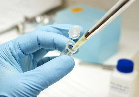
A Bio-Patch Regrows Bone inside the Body

Researchers from the University of Iowa have developed a remarkable new procedure for regenerating missing or damaged bone. It’s called a “bio patch” — and it works by sending bone-producing instructions directly into cells using microscopic particles embedded with DNA.

In experiments, the gene-encoding patch has already regrown bone fully enough to cover skull wounds in test animals. It has also stimulated new growth in human bone marrow stromal cells. Eventually, the patch could be used to repair birth defects involving missing bone around the head or face. It could also help dentists rebuild bone in areas which provides a concrete-like foundation for implants.

To create the bio patch, a research team led by Satheesh Elangovan delivered bone-producing instructions to existing bone cells inside a living body, which allowed those cell to produce the required proteins for more bone production. This was accomplished by using a piece of DNA that encodes for a platelet-derived growth factor called PDGF-B. Previous research relied on repeated applications from the outside, but they proved costly, intensive, and more difficult to replicate with any kind of consistency.

“We delivered the DNA to the cells, so that the cells produce the protein and that’s how the protein is generated to enhance bone regeneration,” explained Aliasger Salem in a statement. “If you deliver just the protein, you have keep delivering it with continuous injections to maintain the dose. With our method, you get local, sustained expression over a prolonged period of time without having to give continued doses of protein.” Salem is a professor in the College of Pharmacy and a co-corresponding author on the paper.

While performing the procedure, the researchers made a collagen scaffold in the actual shape and size of the bone defect. The patch, was loaded with synthetically created plasmids and outfitted with the genetic instructions for building bone did the rest, achieving complete regeneration that matched the shape of what should have been there. This was followed by inserting the scaffold onto the missing area. Four weeks is usually all that it took — growing 44-times more bone and soft tissue in the affected areas compared to just the scaffold alone.

“The delivery mechanism is the scaffold loaded with the plasmid,” Salem says. “When cells migrate into the scaffold, they meet with the plasmid, they take up the plasmid, and they get the encoding to start producing PDGF-B, which enhances bone regeneration.”

The researchers also note that the delivery system is nonviral, meaning that the plasmid is not likely to cause an undesired immune response, and that it’s easier to mass produce, which lowers the cost.

Source: Discovery news


10 medical breakthroughs that sound like science fiction

The news that comes out of research universities and hospitals often sounds too hopeful: Here’s a gene that maybe, could potentially end obesity. This newly discovered protein pathway might sort-of, some day cure cancer. Do any of the thousands of studies published each year really result in a meaningful change in someone’s life?

Here’s your answer: For the eighth consecutive year, the Cleveland Clinic has selected 10 technologies and discoveries that are already making an impact. “We look for innovations that are somewhat disruptive, so a new medication isn’t just a little better, it’s substantially better,” says Dr. Michael Roizen, who headed the panel of 30 medical professionals that selected this year’s finalists. Check out the technology of the future that’s already on our doorstep.

The Bionic Eye

The “Argus II” takes a video signal from a camera built into sunglasses and wirelessly transmits that image to implants in the retinas of people who have lost their vision. Though it’s been available in Europe since 2011, the U.S. Food and Drug Administration (FDA) only approved the eye earlier this year. “This really is like Star Trek technology,” Roizen says.

The system isn’t perfect. It lets a blind person regain basic functions like walking on a sidewalk without stepping off a curb, and distinguishing black from white socks, but only lets you read one giant-sized word at a time on a Kindle. Plus, as the retina itself heals over the implant, the quality of vision decreases. The Argus II is currently only approved for people who have lost their sight from retinal pigmentosis—which affects 1 in 4,000 Americans. But the technology could soon help the more than 1.75 million people who suffer from macular degeneration. (The eyes are the window to the…mind?

The Cancer Gene Fingerprint

Not all cancers are equally lethal—cancer in your prostate means a longer survival rate than a malignancy in your brain, for example. But even prostate cancer comes in multiple flavors ranging from manageable to very bad. By analyzing the mutated genome of a tumor, doctors can now pinpoint whether a cancer is sensitive to a certain chemotherapy, or one that doesn’t respond at all to current treatments. Knowing the subtype might mean jumping directly to a clinical trial that could save your life.

The Seizure Stopper

For the 840,000 epileptics suffering from sudden, uncontrollable seizures, the NeuroPace is like “a defibrillator for your brain,” Roizen says. The system includes sensors implanted in the brain that can spot the first tremors of an oncoming seizure. Then it sends electrical pulses that counteract the brain’s own haywire signals, stopping the seizure in its tracks. Even more impressive: The NeuroPace can be fine-tuned by doctors based on its performance. In the first year it was available, seizure episodes were reduced by an average of 40 percent—but 2 years later, they dropped by 53 percent.

The Hepatitis Cure

Until recently, treatment for hepatitis C fell into the good-but-not-great category, with only around 70 percent of patients being cured. And that was after as much as 48 weeks of a strict anti-viral drug regimen, including injections of interferon—which causes a number of debilitating side effects. But the new drug Sofosbuvir is a much more potent killer of hep C, with success in as many as 95 percent of patients. Even more, the medication only has to be administered for 12 weeks, sans interferon injections.

The Anesthesiologist’s iPad

Surgeons may get more glory, but anesthesiologists probably play the most vital role in keeping you alive during surgery. They’re the last face you see before you’re put into a medicated sleep so deep you don’t even notice that your body is being peeled open. Between keeping track of your heart rate, breathing, and brain functions, an anesthesiologist also needs to be familiar with the ins and outs of the procedure so they can adjust sedatives and painkillers—without causing complications. 

The new “perioperative information management systems” include software on touchscreen-enabled computers that can warn doctors if things are going south, keep track of the surgeon’s workflows, and document every step of the procedure. All are essential when surgeries last up to 16 hours and docs need to pass the reins to a fresh pair of eyes.

The Fecal Transplant

The idea of taking someone else’s poop and giving it a new home in your own colon may sound repulsive, but the treatment has proven remarkably effective in curing infections of C. difficile—a nasty bacteria that kills 15,000 people each year. Take heart: The digested food waste in feces isn’t itself the cure. You’re simply gaining some of the helpful bacteria living in the donor’s gut—like a farmer choosing the hardiest crops to seed next year’s fields.

“The bacteria produce proteins that are involved in a lot more diseases than we realized,” says Roizen. Still grossed out? Researchers in Canada have developed a method to deliver just the bacteria—no feces—via an oral pill, skipping the need for a poo enema.

The Heart-Saving Hormone

Around one in four people who are hospitalized for heart failure don’t last much longer than a year. But a new drug called Serelaxin has upped the odds of survival by as much as 37 percent, according to a University of California, San Francisco study. It’s a synthetic version of the hormone relaxin, which is produced by pregnant women to help with the increased stress carrying a fetus places on the heart. “It not only opens up your blood vessels to supply your organs oxygen, but it has anti-inflammatory properties,” Roizen says. Serelaxin’s life-saving potential is profound enough that in June, the FDA dubbed it a “breakthrough therapy,” putting it on a faster track for approval in hospitals.

The Robot Doctor

If you’re undergoing a colonoscopy, you’ll want something to take the edge off (for obvious reasons). But even a light sedative to help you snooze while doctors spelunk your butt requires the presence of an anesthesiologist—which translates to $1 billion in additional medical expenses, according to a study in the Journal of the American Medical Association.

Enter the Sedasys: a computer with an attachment on the IV that meters out the correct amount of sedative and monitors vitals. It even includes an earpiece to wake patients up if necessary. That allows docs to administer “light to moderate” sedation on their own, with a single anesthesiologist supervising multiple patients. “If Michael Jackson’s doctor had this and knew how to use it, then Michael Jackson would still be alive today,” says Roizen.

The Better Heart-Attack Risk Test

Today you get a cholesterol test to assess your risk of heart attack, but soon you’ll be more worried about your trimethylamine N-oxide (TMAO) levels. Why? People with the highest levels of TMAO in their blood have 2.5 times the risk of a heart attack compared to those with the lowest levels, according to a recent study in the New England Journal of Medicine. TMAO is a compound produced by intestine bacteria—yep, the same ones involved in fecal transplants—after you eat choline, which is found in eggs, red meat, and dairy.

Once in your bloodstream, TMAO accelerates the process of cholesterol forming into plaques in your arteries. “We’re learning why red meat is hazardous, and what could be done to avoid that hazard,” Roizen says. Beyond simply avoiding red meat, preventive steps could include probiotics or medications that pinch off TMAO-producing pathways.

The Precision-Guided Cancer Treatment

The difficult goal in any cancer treatment is to kill the tumor while leaving healthy cells alone. Recently, a better understanding of what makes cancer cells tick has allowed scientists to develop a class of drugs that pinpoint a weakness in cancer’s uncontrolled growth. For example, in lymphomas and leukemias, scientists have determined that the growth is controlled by a protein called Bruton’s tyrosine kinase (BTK). After years of experimentation, doctors developed a new drug called Ibrutinib that blocks BTK

Source: inagist


Surgeon Wins Award For 95p Invention

Dr Hossien spent over six months developing his invention A heart surgeon at a Swansea hospital has won an award for an invention that cost him 95p to create. Morriston hospital doctor Abdull razak Hossien made his surgery training simulator out of a sweet tin. The portable device can be used anywhere and is now being manufactured for use around the world

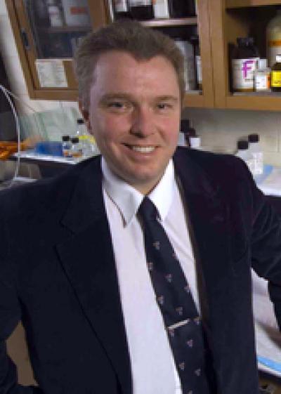
Dr Hossien spent over six months developing his invention

A heart surgeon at a Swansea hospital has won an award for an invention that cost him 95p to create.

Morriston hospital doctor Abdullrazak Hossien made his surgery training simulator out of a sweet tin.

The portable device can be used anywhere and is now being manufactured for use around the world.

It is used in training for surgery of the aortic root, which carries blood from the left side of the heart to the arteries of the limbs and organs.

Dr Hossien created his training device for a competition run as part of the European Association for Cardiothoracic Surgery (EACTS) Conference 2013 in Vienna.

He said: “Thomas Edison said that to invent you need a good imagination and a pile of junk.

“I designed a portable trainer, which you can keep in your pocket. You can practise on the train, on an airplane, at home, wherever you are.

“I developed it from a sweet tin that can be fixed to a table, and created an aorta using synthetic material. It cost me around 95p.

“I accompanied this simulator with a multimedia DVD [with] guidelines that trainees can apply to any procedure on the aortic root. They can progress from the simplest procedure to the most complex as they develop.”

Dr Hossien said trainees using the simulator would be completely familiar with the procedures by the time they came to carry out supervised aortic root surgery on patients.

He added: “They will have mastered the procedure before they operate on the patient.”

Garage workshop

Dr Hossien said that at the same time “qualified surgeons and any doctor with an interest in the specialty can improve their skills”.

The prototype simulator was made from a sweet tin

For the EACTS award, candidates were challenged to create a low-tech training simulator for aortic root surgery.

These were judged by a panel of eight top surgeons from Europe and the USA.

Dr Hossien was eventually declared joint winner along with a candidate from Italy.

His simulator will be manufactured for worldwide use by award sponsor Ethicon, which develops innovative surgical products.

Dr Hossien turned the garage of his Swansea home into a workshop to develop the aortic root simulator.

“I spent six or seven months on it. I would forget to eat and to drink sometimes because I was thinking about it so much.

“I would like to thank my wife and daughter who supported me and gave me the time I needed to develop this.”

He is donating his share of the 3,000 euro (£2,600) first prize from the EACTS award to the Syrian humanitarian relief appeal.

Dr Hossien is senior clinical research fellow in the cardiothoracic department at Morriston Hospital.

Mr Saeed Ashraf, consultant cardiothoracic surgeon and honorary senior lecturer at Swansea University said: “Dr Hossien is a very talented academic surgeon with an excellent pair of surgical hands.”

Source: 24all news


New Drug Combination Causes Cancer To ‘Eat’ themselves

Turning cancerous cells into cannibalistic cells may be an oncologists best bet for treating a wide variety of tumors. In a study from the Virginia Commonwealth University, researchers have determined that a new drug combination can cause cancer cells to “eat” themselves without harming surrounding tissue. The treatment was shown to be effective in cancers of the colon, liver, lung, breast, brain, and kidney.

Published in the journal Molecular Pharmacology, the study sought to determine whether a combination of the drugs sorafenib and regorafenib can combine with so-called PI3K/AKT inhibitors to achieve a broader, more pronounced effect. Co-author Andrew Poklepovic said in a press release that although further research is necessary, the current findings set the stage for a promising phase 1 clinical trial.

“It is still too premature to estimate when a clinical trial will open to further test this drug combination therapy, but we are now in the planning phase and encouraged by the results of these laboratory experiments,” he told reporters.  “We are also encouraged by the fact that the drugs used in this therapy are either already approved by the FDA to treat certain cancers or are currently being investigated in other clinical trials.”

According to the researchers, sorafenib and regorafenib attacks the tumor growth by blocking the production of enzymes called kinases, which play a crucial role in the proliferation of cancerous cells. Essentially, the new drug combination broadens this effect by adding another class of kinases inhibitors called PI3K/AKT inhibitors. As a result, many cancer cells “starve,” and the tumor growth can no longer be sustained.

“We know that there are certain cellular processes that are frequently dysregulated in cancers and important to cell proliferation and survival, but if you shut down one, then cells can often compensate by relying on another,” said senior author Paul Dent of the Virginia Commonwealth University Massey Cancer Center. “We are blocking several of these survival pathways, and the cancer cells are literally digesting themselves in an effort to stay alive.”

The study represents the latest attempt to identify a new, reliable method of action for cancer drugs. In a similar research effort published earlier this year, scientists at Oregon State University described how a new type of nanotechnology can improve the efficacy of cytotoxic therapies by heating up cancerous cells. Hopefully, both studies will help oncologists and pharmaceutical researchers develop novel ways to treat the disease that currently kills over 500,000 Americans each year.

Source: Medical Daily


Cancer vaccine developed to boost lifespan of patients

Russian scientists have developed a vaccine for the treatment of cancer that can increase the patient’s lifespan more than two-fold, ITAR-TASS reported on Wednesday.

The vaccine, developed at the Institute of Clinical Immunology in the Siberian branch of the Russian Academy of Medical Sciences in Novosibirsk, has already successfully passed clinical tests, said institute’s director Vladimir Kozlov.

Currently, it is being administered to patients by injection at the third and fourth stages of cancer.

”We are deriving dendritic cells from the human body and loading them with tumor antigens,” Kozlov said.

“Dendritic cells process them, then we inject the cells into the patient and they start working in the body evoking a strong immune response. That is, they are actively fighting the tumor.”

The institute’s creation “is not a classical vaccine, which is the means to prevent the illness”, Kozlov said. “In fact, it is a cell therapy.”

The vaccine is counteracting several types of cancer – colorectal (bowel) cancer, breast cancer and prostate cancer – and the institute is ready to start its industrial production, Kozlov added.

However, the scientist added that other types of therapy were to be used as well, such as chemotherapy and radiation therapy.

Source: Khaleej Times


Chinese vaccine to save Asian children from encephalitis gets WHO nod

China’s first vaccine to save children in Asia from deadly Japanese encephalitis has been approved for global use by the World Health Organization.

The vaccine, manufactured by the Chengdu Institute of Biological Products, has received WHO prequalification, which means it meets international standards for quality, safety and efficacy.

According to China Daily, WHO Director-General Margaret Chan said that the move from China, which is producing vaccines up to WHO standards, is a welcome development, both in the fight to protect children in developing countries from the virus and in the future availability of vaccines more generally.

She said that there is a huge potential for vaccine manufacture in China, adding that she hopes to see more Chinese vaccines get WHO prequalification. The whole world will benefit.

The GAVI Alliance, a public-private global health partnership committed to saving children and increasing access to immunization in poor countries, said it is preparing to make funding available for the vaccine, the report said.

CEO Seth Berkley said that the Chinese vaccine industry has huge potential to benefit children in the poorest countries by offering secure, predictable supply at affordable prices, the report added.

The GAVI Alliance brings together governments, the WHO, UNICEF, the World Bank, vaccine industries in industrialized and developing countries, research and technical agencies and civil societies and the Bill and Melinda Gates Foundation and other private philanthropists.

Japanese encephalitis is a vicious illness that strikes quickly and usually has a devastating impact on children and their families, Berkley said.

Source: http://bit.ly/HGM7S2


Lasers could potentially cure Alzheimer’s and Parkinson’s

Researchers at South Africa’s Council for Scientific and Industrial Research (CSIR) have unveiled the world’s first digital laser

Lasers (photo therapy) might someday be the cure for brain diseases such as Alzheimer’s, Parkinson’s or Creutzfeldt-Jakob disease. Researchers at Chalmers University of Technology in Sweden, together with researchers at the Polish Wroclaw University of Technology, have made a discovery that may lead to the curing of diseases such as Alzheimer’s, Parkinson’s and Creutzfeldt-Jakob disease (the so called mad cow disease) through photo therapy. You can check out the abstract of the latest study, “Multiphoton absorption in amyloid protein fibers” published in the November 3, 2013 issue of Nature Photonics.

Lasers might be the cure for brain diseases such as Alzheimer’s and Parkinson’s. Zap your Alzheimer’s or Parkinson’s with a laser light instead of a drug or invasive surgery since a properly functioning protein is optically invisible to high power laser light, and toxic amyloid is responsible for brain diseases. A special laser zap on your Alzheimer’s or Parkinson’s could be the answer. These diseases might potentially be cured using lasers in photo therapies. Since the different amyloids have the same structure, perhaps they can be zapped by certain types of laser light using a process known as photo therapy.

After all, how many patients would prefer photo therapy to the side effects of drugs that may not work or other procedures that are more invasive?

Researchers at Chalmers University of Technology in Sweden, together with researchers at the Polish Wroclaw University of Technology, have made a discovery that may lead to the curing of diseases such as Alzheimer’s, Parkinson’s and Creutzfeldt-Jakob disease (the so called mad cow disease) through photo therapy. The researchers discovered, as they show in the journal Nature Photonics that it is possible to distinguish aggregations of the proteins, believed to cause the diseases, from the the well-functioning proteins in the body by using multi-photon laser technique.

“Nobody has talked about using only light to treat these diseases until now. This is a totally new approach and we believe that this might become a breakthrough in the research of diseases such as Alzheimer’s, Parkinson’s and Creutzfeldt-Jakob disease. We have found a totally new way of discovering these structures using just laser light”, says Piotr Hanczyc at Chalmers University of Technology, according to the November 3, 2013 news release, “Lasers might be the cure for brain diseases such as Alzheimer’s and Parkinson’s.”

If the protein aggregates are removed, the disease is in principle cured. The problem until now has been to detect and remove the aggregates

The researchers now harbor high hopes that photo acoustic therapy, which is already used for tomography, may be used to remove the malfunctioning proteins. Today amyloid protein aggregates are treated with chemicals, both for detection as well as removal. These chemicals are highly toxic and harmful for those treated.

With multi photon laser the chemical treatment would be unnecessary. Nor would surgery be necessary for removing of aggregates. Due to this discovery it might, thus, be possible to remove the harmful protein without touching the surrounding tissue.

These diseases arise when amyloid beta protein are aggregated in large doses so they start to inhibit proper cellular processes. Different proteins create different kinds of amyloids, but they generally have the same structure. This makes them different from the well-functioning proteins in the body, which can now be shown by multi photon laser technique. You may also wish to check out the site of the Foundation for Polish Science Welcome Grant, European Research Council or Chalmers University of Technology.

Here’s how the process works

Fibrillization of peptides leads to the formation of amyloid fibers, which, when in large aggregates, are responsible for diseases such as Alzheimer’s and Parkinson’s. In the latest study, researchers show that amyloids have strong nonlinear optical absorption, which is not present in native non-fibrillized protein. Z-scan and pump–probe experiments indicate that insulin and lysozyme β-amyloids, as well as α-synuclein fibres, exhibit either two-photon, three-photon or higher multiphoton absorption processes, depending on the wavelength of light.

The researchers proposed that the enhanced multiphoton absorption is due to a cooperative mechanism involving through-space dipolar coupling between excited states of aromatic amino acids densely packed in the fibrous structures. This finding will provide the opportunity to develop nonlinear optical techniques to detect and study amyloid structures and also suggests that new protein-based materials with sizable multiphoton absorption could be designed for specific applications in nanotechnology, photonics and optoelectronics.

It’s a possibility that someday shining a laser light on someone might be able to cure Parkinson’s and Alzheimer’s or Creutzfeldt-Jakob disease. The idea is that if the different amyloids have the same structure, perhaps they can all be zapped by certain types of laser light.

Source: http://exm.nr/1cBmAYK


Scientists regenerate fully functional tear and saliva glands

A research group at Tokyo University of Science has successfully regenerated fully functional bioengineered salivary and lacrimal (tear) glands. The results signify a substantial advance in the development of next generation organ replacement regenerative therapies. The results are published in the scientific journal Nature Communications.

Organ replacement regenerative therapy has been proposed as having the potential to enable the replacement of organs that have been damaged by disease, injury or aging. The research group led by Professor Takashi Tsuji (Professor in the Research Institute for Science and Technology, Tokyo University of Science, and Director of Organ Technologies Inc.) has provided a proof-of-concept for bioengineered organ replacement as the next step for regenerative therapy.

For the salivary glands, Tsuji’s group reports the fully functional regeneration of a salivary gland that reproduces the morphogenesis induced by reciprocal epithelial and mesenchymal interactions through the orthotopic transplantation of a bioengineered salivary gland germ as a regenerative organ replacement therapy. The bioengineered germ developed into a mature gland through acinar formations with the myoepithelium and innervation. The bioengineered submandibular gland produced saliva in response to the administration of pilocarpine and gustatory stimulation by citrate, protected against oral bacterial infection and restored normal swallowing in a salivary gland defect mouse model. Thus, this study provides a proof-of-concept for bioengineered salivary gland regeneration as a potential treatment for xerostomia.

For the lacrimal (tear) glands, Tsuji’s group reports the successful orthotopic transplantation of a bioengineered lacrimal gland germ into an adult extra-orbital lacrimal gland defect model mouse, which mimics the corneal epithelial damage caused by lacrimal gland dysfunction. The bioengineered lacrimal gland germ and harderian gland germ both developed in vivo and achieved sufficient physiological functionality, including tear production in response to nervous stimulation and ocular surface protection. This study demonstrates the potential for bioengineered organ replacement to functionally restore the lacrimal gland.

Source:


Sputum test may not hold key to TB eradication: study

Sputum test for tuberculosis works better than the commonly-used, 125-year-old microscope method, but did not reduce illness in a southern African trial, a study said today.

While the new test, dubbed Xpert MTB/RIF, diagnosed tuberculosis (TB) quicker and got people onto treatment sooner, it did not seem to change their long-term prognosis, said the study in The Lancet medical journal.

“Earlier diagnosis by the Xpert test did not reduce overall severity of TB-related illness,” study leader Keertan Dheda from the University of Cape Town’s department of medicine said in a statement.

Xpert was put to the test in a trial in South Africa, Zimbabwe, Zambia and Tanzania — countries with high rates of TB — from April 2011 to March 2012.

A total of 744 people with suspected TB symptoms were randomly assigned to undergo the Xpert test and another 758 the traditional method of using a microscope to examine lung matter for TB bacteria.

Called smear microscopy, this method is often combined with a chest x-ray, and is estimated to miss about 40 to 60 per cent of TB cases.

On top of this, about 40 per cent of people who do test positive never return to the clinic for their test results and treatment — hence the need for a faster diagnostic test like Xpert, which can show a result within two hours.

Xpert was endorsed by the World Health Organisation (WHO) in 2011 for people with suspected multi-drug-resistant TB, and TB complicated by simultaneous HIV infection.

In the southern African trial, 185 of the subjects tested positive in the Xpert test and 182 in the other.

They were examined again at two months and six months after starting treatment, using a scoring system that measured their quality of life and signs and symptoms of TB, said the study authors.

“Despite a longer delay to treatment in the microscopy group, no difference in the severity of TB-related illness, which correlates well with longer term prognosis, was noted after two months and six months between the two groups,” the authors said.

By the end of the study, eight per cent of patients in both groups had died.

“The potential long-term effect of this test is probably overestimated,” concluded the study.

The findings would be of major interest to policymakers “because the costs of rolling out Xpert MTB/RIF are very high”, Christian Wejse from the public health department of Denmark’s Aarhus University wrote in a comment also carried by The Lancet.

Reversing the incidence of TB is one of the UN Millennium Development Goals for 2015, but the disease remains a major cause of illness and death.

Untreated, TB kills about half the people it infects.

In 2012, 8.6 million people fell ill with the lung disease and 1.3 million died from it, according to the WHO.

Over 95 per cent of TB deaths occur in low- and middle-income countries, and it is among the top three causes of death for women aged 15 to 44.

Source:


Elusive Secret of HIV Long-Term Immunity

Discovery offers hope of new, shorter HIV treatment if drugs are started right away

Scientists have discovered a critical new clue about why some people are able to control the HIV virus long term without taking antiviral drugs. The finding may be useful in shortening drug treatment for everyone else with HIV.

These rare individuals who do not require medicine have an extra helping of a certain type of immune protein that blocks HIV from spreading within the body by turning it into an impotent wimp, Northwestern Medicine® scientists report. The new finding comes from analyzing cells from these rare individuals and HIV in the lab.

Scientists have been trying to solve the mystery of why 1 percent of people with HIV — called “controllers” — have enduring control of the virus without medications, in some cases for life. The controllers’ early defense against HIV is quickly extinguished by the virus, so how do they have long-term immunity? The Northwestern discovery represents what scientists have long sought: a second line of defense deep in the immune system backing up the short-lived early defense.

This discovery suggests a novel approach involving much earlier treatment that could potentially make every HIV-infected person into a controller for the long term by protecting the reserves of this defensive immune protein. The goal would be for them to eventually be free from anti-retroviral drugs.

Currently most HIV patients need to take powerful anti-retroviral drugs every single day for life. If the medicines are stopped, the virus quickly reactivates to harmful levels even after years of treatment.

“Preserving and even increasing this defense in cells may make more HIV-infected persons into controllers and prevent HIV from rebounding to high and damaging levels when anti-HIV medications are stopped,” said Richard D’Aquila, M.D., the director of the Northwestern HIV Translational Research Center. He is the senior author of the study, which will be published Oct. 16 in the journal PLOS ONE.

D’Aquila also is the Howard Taylor Ricketts Professor of Medicine at Northwestern University Feinberg School of Medicine and a physician at Northwestern Memorial Hospital.

D’Aquila and colleagues now are working to develop a medicine that would boost this defensive immune protein called APOBEC3G, or A3 for short. 

The Missing Second Defensive Line

Much is known about how the immune system of controllers initially fights the virus. But HIV quickly escapes from that first line of defense by mutating and evading the adaptive immune system. How these individuals control HIV long term without medications to keep from developing AIDS has been under study by many researchers? It seemed there must be a second defensive line in the immune system.

Turning HIV Into a Wimp

In the new study, D’Aquila and his team have found that controllers, long after they have acquired HIV, have a more abundant supply of the critical immune protein A3 in specific white blood cells called resting memory T cells. This is where the virus lies silently in an inactive form and roars back when anti-retroviral drugs are stopped. In controllers, though, their bounty of A3 means that any new HIV made from those cells inherits a helping of A3, which turns the new viruses into harmless wimps that can’t infect other cells.

You Can’t Fool A3

The feisty A3 is a critical part of the newly characterized intrinsic immune system, and it resides in many cells of the immune system including resting T cells. Unlike the adaptive immune system, which fails to recognize the virus once it mutates its pieces, the intrinsic immune system can’t be fooled.

“The intrinsic immune system recognizes the basic guts of the virus — the nucleic acids — that HIV can’t change and then damages those nucleic acids,” D’Aquila said.

D’Aquila theorizes that the controllers’ first line of defense slows down the ability of HIV to destroy all the A3.

“Perhaps starting anti-HIV drugs very soon after HIV is caught, rather than the current practice of waiting until later to start, would work like the controllers’ first line of defense,” D’Aquila suggested. “If we preserve A3, it could minimize HIV’s spread through the body as this protein seems to do in controllers.”

Otherwise, D’Aquila theorizes, all reserves of the protein are wiped out if HIV replicates unchecked for several months.

See more at: