Smart phones to diagnose diseases

Not just helping us with road directions, finding favorite food joint, photo sharing and chat sessions, smart phones would soon diagnose diseases for us in real time.

Researchers from University of Houston are developing a disease diagnostic system that offers results that could be read using only a smart phone and a Rs.1,200 lens attachment.

This new device relies on specific chemical interactions that form between something that causes a disease – a virus or bacteria, for example – and a molecule that bonds with that one thing only, like a disease-fighting antibody.

A bond that forms between a strep bacteria and an antibody that interacts only with strep, for instance, can support an iron-clad diagnosis.

“The trick is finding a way to detect these chemical interactions quickly, cheaply and easily. The device involves a simple glass slide and a thin film of gold with thousands of holes poked in it,” explained Jiming Bao, assistant professor of electrical and computer engineering at University of Houston.

The device starts with a standard slide covered in a light-sensitive material known as a photoresist. It uses a laser to create a series of interference fringes – basically lines – on the slide, and then rotates it 90 degrees and creates another series of interference fringes.

The intersections of these two sets of lines creates a fishnet pattern of UV exposure on the photo-resist. The photo-resist is then developed and washed away.

While most of the slide is then cleared, the spots surrounded by intersecting laser lines – the ‘holes’ in the fishnet – remain covered, basically forming pillars of photo-resist.

The end result is a glass slide covered by a film of gold with ordered rows and columns of transparent holes where light can pass through. These holes are key to the system. The device diagnoses an illness by blocking the light with a disease-antibody bond – plus a few additional ingredients.

Here is where the smart phone comes in. “One of the advantages of this system is that the results can be read with simple tools,” said Richard Willson, Huffington-Woestemeyer professor of chemical and biomolecular engineering.

A basic microscope used in elementary school classrooms provides enough light and magnification to show whether the holes are blocked.

With a few small tweaks, a similar reading could almost certainly be made with a phone’s camera, flash and an attachable lens.

This system, then, promises readouts that are affordable and easy to interpret. There are some technical hurdles to clear before the system can be rolled out, Willson noted. One of the biggest challenges is finding a way to drive the bacteria and viruses in the sample down to the surface of the slide to ensure the most accurate results.

But if those problems are overcome, the system would be an excellent tool for health care providers in the field, said the research published in the journal ACS Photonics.


Silk screws used to repair fractures

silk_screws

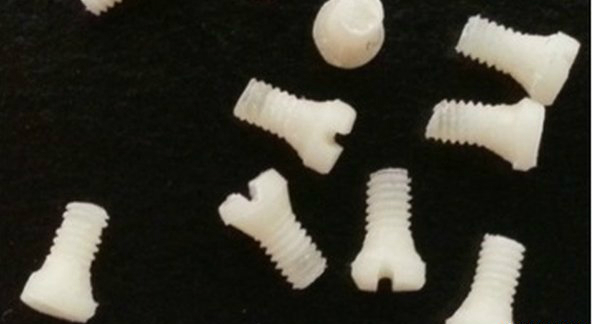
Screws made from 100% silk have been used to repair broken bones in research that could transform surgery.

US scientists say metal fixtures can potentially be replaced with plates and screws made from the natural fibre, which will eventually dissolve in the body.

So far the technique has only been tested on rodents. Silk was once used to make sutures, but more recently has been used in modern medical implants.

In the new research, a team of medical engineers at Tufts University, Massachusetts, made screws from medical grade silk using specially designed moulds. The silk material can be cut to different sizes on a machine.

The screws were implanted into the hind limbs of rats, where they functioned successfully for four to eight weeks.

By the end of the study, the silk had started to dissolve. The low stiffness of silk, which is similar to that of bone, and its ability to break down in the body, make it a promising bioengineering material compared with traditional metal plates and screws, the researchers say.

Lead researcher Dr David Kaplan said “The future is very exciting. We envision a whole set of orthopaedic devices for repair based on this – from plates and screws to almost any kind of device you can think of where you don’t want hardware left in the body.”

He added: “They don’t interfere with X-rays, they don’t set off alarms and they don’t cause sensitivity to cold.”

Recently, German researchers coated silicone breast implants with a thin layer of bioengineered silk proteins.

Preclinical studies suggest the coating reduces or prevents painful reactions.

The new research is published in the journal, Nature Communications.

Source: BBC news


3D-printed heart aids life-saving surgery on US baby

3d-printed-splint-family-13

A newly developed 3D-printed heart has helped doctors perform a life-saving heart surgery on a 14-month old infant in the US.

Researchers from the University of Louisville and Kosair Children’s Hospital created a 3D printed model of the organ 1.5 times its actual size that helped the surgeons to prepare for the surgery.

Built in three pieces using a flexible filament, the printing reportedly took around 20 hours and cost USD 600.

Roland Lian Cung Bawi of Owensboro, Kentucky, was born with four congenital heart defects and his doctors were looking for greater insights into his condition prior to a February 10 operation.

Philip Dydynski, chief of radiology at Kosair Children’s Hospital wondered if a 3D model of the child’s heart could be constructed using a template created by images from a CT scan to allow doctors to better plan and prepare for his surgery.

The result was a model heart 1.5 times the size of the child’s.

Once the model was built, Erle Austin III, cardiothoracic surgeon at Louisville, was able to develop a surgical plan and complete the heart repair with only one operation.

“I found the model to be a game changer in planning to do surgery on a complex congenital heart defect,” he said.

Roland was released from Kosair Children’s Hospital on February 14. His prognosis is good, doctors said.

Source: zee news


New robotic surgical institute opens in the Dominican Republic

This week the Metropolitan Hospital of Santiago (HOMS), in the Dominican Republic, opened the first robotic surgery institute in the Caribbean. Named for Dr. David Samadi, a pioneer in robotic prostate surgery and member of the Fox News Medical A-Team, it is sure to draw patients from the United States as well from the Caribbean and South America.

Nearly one million Americans seek health care outside the U.S. every year. Medical tourism is increasingly popular in the Caribbean both in terms of its high quality, cost savings of up to 75 percent, and of course, you can’t beat the beautiful surroundings. The Dominican Republic has long been a tourist destination known for its inexpensive plastic surgery and dental procedures.

Now you can add to that the state-of-the-art Davinci robot which is like at avatar where the surgeon uses precision robotic arms to perform delicate procedures. Recovery with this kind of surgery is remarkable – you can get in and out of the hospital in a day. At a time when America is debating its health care future, our latest and greatest technology is still being exported.

Dominican Republic President Danilo Medina is opening the new facility along with Dr. Raphael Sanchez Espanol, CEO of HOMS hospital and, himself, a renowned surgeon.

“You’re going to change the lives of many, many people out there, and to be part of the first robotic institute in the Caribbean, this is a huge blessing and for the humanitarian part… for us to be part of this we’re very proud,” said Dr. David Samadi, chairman of urology and chief of robotic surgery at Lenox Hill Hospital in New York City.

The institute will include treatment of prostate, kidney, gynecologic and surgical cases. Doctors operating in the Dominican Republic will now be able to use techniques developed by Samadi, who performed the country’s first procedure, to treat prostate cancer as well as other delicate surgeries in a safer, more effective manner with minimal blood loss.

“Big decisions have transformed into small decisions,” said Dr. Espanol. “I like to say, with less trauma – the less trauma facilitates an amazing recovery of the sick.”

“This is a huge step in this country and it’s going to bring a lot of patients from the entire Caribbean to this hospital,” Samadi said.

And beyond the Caribbean, with more and more regulations and restrictions coming our way on American soil, many patients will look elsewhere for their surgeries. The new David Samadi Robotic Institute is one of the places they will be looking.

Source: Fox news


Delhi gets Asia’s first ‘Silent’ MRI system

With a vision of laying as much importance on ‘patient-comfort’ as on the actual medical services provided to them, Mahajan Imaging, north India’s leading chain of high-end medical imaging centres, on Tuesday claimed to have installed Asia’s first ‘Silent’ MRI system in a ‘unique, non-intimidating and non-medical environment’ at its new diagnostic centre in Defence Colony here.

According to Mahajan Imaging, the MR750w MRI system by GE Healthcare has the world’s most advanced imaging technology that enables radiologists to perform MRI scans “without any sound”.

According to GE Healthcare, its revolutionary Silent Scan technology is designed to reduce MR scanner noise to near ambient (background) sound levels and thus improve a patient’s MR exam experience. Conventional MR scanners can generate noise in excess of 110 dBA (decibels) levels, roughly equivalent to rock concerts.

Dr Harsh Mahajan, chief radiologist at Mahajan Imaging, said, “We have installed Asia’s first truly silent MRI system. Till now, MRI scans were associated with a lot of noise – almost as much as a jet engine or rock concert, which led to stress and discomfort for patients. Today, we are able to do most brain scans without any noise at all! This, in addition to the obvious benefit of comfort to the patient, also enables us to do higher quality scans since patients do not move and are more cooperative during scans. This eventually leads to better images and hence a better diagnosis.”

Dr Mahajan said that soon the ‘silent’ technology would be extrapolated to other parts of the body, which would lead to a paradigm shift in MRI scanning.

“The development of this revolutionary Silent Scan MRI Technology is in keeping with GE’s mission of humanizing its medical technology,” said Karthik Kuppusamy, senior director of MR Imaging at GE Healthcare South Asia.

“This machine is also testament of India’s product development capabilities since it was co-developed by our engineers in Bangalore. We used proprietary high-fidelity gradient and ultra-fast RF system electronics and eliminated the sound at the source,” he added.

“It is amazing that there is no drop in image quality,” said Dr Raju Sharma, professor of radiology at the All India Institute of Medical Sciences (AIIMS), New Delhi. “I congratulate GE for developing a clinical system that can perform MRI scans without any sound – this is a dream come true for radiologists all over the world.”

Dr Sharma explained that having a noise-free environment makes the patients very comfortable. This improves utilisation of the MRI machine since scans do not need to be interrupted or repeated frequently.

Additionally, Mahajan Imaging has taken patient comfort a step further by giving both, their MRI room and CT room very unique and aesthetically pleasing interiors. While the MRI room bears the theme — Genesis-Hope-Healing, depicted using a 33 feet long painting of three Greek Goddesses, the CT scan room gives the impression of being underwater.

“The idea is to create an environment of healing,” said Ritu Mahajan, executive director of Mahajan Imaging and the person responsible for designing the facility. “We decided to push the boundaries of what is possible in an MRI room and used silk on the walls and ceiling to add warmth, and matching self-levelling epoxy flooring. Additionally, an artist from Albania was approached to make a 33 feet long painting which depicts Genesis, Hope and Healing, according to Greek mythology.”

“The room housing the CT scan machine in the diagnostic centre has been hand-painted by a young artist to give the impression of being underwater, something that is very popular with younger patients,” she said.

“Since an MRI machine has a very strong magnetic field, and is affected even by extremely small changes in radiofrequency waves, we decided to go with several small specially manufactured LED lights to light up the MRI room,” said Dominique Taffin, senior design architect from GE Healthcare who helped execute the MRI room interiors at the centre.

“The positive effect of one’s surroundings on one’s health cannot be underplayed,” said Dr S S Kale, professor of neurosurgery at AIIMS, New Delhi. “What Dr Mahajan and his team have done is truly unique – every room has a different theme! The environment at the centre sets the patient at ease – a very important component of providing healthcare from a holistic point of view.”

Niharika Dutt, a 22-year old healthcare professional, has had several MRI scans done earlier because of some illness, but feels that this machine is different. “I have had MRI scans done many times because of repeated dizziness and migraine,” said Dutt, “but I have never seen a room this pretty! I had seen photographs of this room online and felt that they were edited – but I later realised that Mahajan Imaging has actually made their room like this. The ambience, in addition to the quietness of the machine, is definitely going to make any patient forget about their disease for some time and make the harrowing experience of the MRI better.”

In addition to the advanced MRI and CT technology, the new centre boasts of the world’s best Mammography machine, X-Ray system, ultrasounds, cardiac scanners, dental CT and DEXA machine – providing all radiology services under one roof.

Source : India Medical Times


Virtual arm eases phantom limb pain

Doctors have devised a new way to treat amputees with phantom limb pain.

Using computer-generated augmented reality, the patient can see and move a virtual arm controlled by their stump.

Electric signals from the muscles in the amputated limb “talk” to the computer, allowing real-time movement.

Amputee Ture Johanson says his pain has reduced dramatically thanks to the new computer program, which he now uses regularly in his home.

He now has periods when he is free of pain and he is no longer woken at night by intense periods of pain.

Mr Johanson, who is 73 and lives in Sweden, lost half of his right arm in a car accident 48 years ago.

After a below-elbow amputation he faced daily pain and discomfort emanating from his now missing arm and hand.

Over the decades he has tried numerous therapies, including hypnosis, to no avail.

Within weeks of starting on the augmented reality treatment in Max Ortiz Catalan’s clinic at Chalmers University of Technology, his pain has now eased.

“The pain is much less now. I still have it often but it is shorter, for only a few seconds where before it was for minutes.

“And I now feel it only in my little finger and the top of my ring finger. Before it was from my wrist to my little finger.”

Mr Johanson says he has noticed other benefits too. He now perceives his phantom hand to be in a resting, relaxed position rather than a clenched fist.

“Can you imagine? For 48 years my hand was in a fist but after some weeks with this training I found that it was different. It was relaxed. It had opened.”

Mr Johanson has also learned to control the movements of his phantom hand even when he is not wired up to the computer or watching the virtual limb.

Max Ortiz Catalan, the brains behind the new treatment, says giving the muscles a work-out while being able to watch the actions carried out may be key to the therapy.

“The motor areas in the brain needed for movement of the amputated arm are reactivated, and the patient obtains visual feedback that tricks the brain into believing there is an arm executing such motor commands. He experiences himself as a whole, with the amputated arm back in place.”

He says it could also be used as a rehabilitation aid for people who have had a stroke or those with spinal cord injuries.

Source: BBC news


New ‘painless’ treatment to repair teeth

A novel “regenerative” technique to repair infected teeth – claimed to be painless and cheaper than the traditional root canal treatment – has been developed by doctors at the All India Institute of Medical Sciences (AIIMS) in New Delhi, says IANS.

Termed as “SealBio”, the technique uses body’s own stem cells and eliminates the need for cumbersome root canal fillings.

Developed by doctors Naseem Shah and Ajay Logani at the Department of Conservative Dentistry and Endodontics, it has obtained an Australian patent, while an application with the US Patents office is under process.

Root canal treatment demands rigorous training, clinical skills and several cumbersome sittings with dentist. It involves thorough cleaning and shaping followed by filling of the entire root canal with one of several sealer cements.

The AIIMS technique is claimed to be the first that dispenses with the need for root canal filling. Instead of filling the root canal with artificial materials that may pose bio-compatibility problems, it makes use of regenerative potential of stem cells and growth factors available at the root of the teeth. Stem cells act as a repair system for the body capable of replenishing adult tissues.

In case of this technique, the stem cells at the root of the decayed teeth are stimulated to induce regeneration and deposition of a natural tissue barrier (seal) to fill up the root canal in just one sitting.

In other words, a “biological seal” is achieved at the root canal rather than attempting to seal it with artificial filling materials with all its drawbacks, the doctors say. The root canal is restored to health by gradual build up of tissue by stem cells over a period, extending from a few weeks to some months.

The AIIMS doctors say that this treatment simplifies the whole procedure with minimal use of equipment, less time and cost of treatment.

“Shah has been able to successfully carry out this new procedure in dozens of patients and the 4-5 years follow up results have been very encouraging,” Seyed Hasnain, a professor at the Kusuma School of Biological Sciences of the Indian Institute of Technology, Delhi, told IANS on phone. “In my opinion this is a path breaking innovation, a game changer,” Hasnain said.

He said that the success achieved by AIIMS doctors could trigger research in regenerative techniques in other clinical situations in dental science.

Source: The Free Press


A Japanese iPhone could bring you the Food Smells

Have you ever wished that your iPhone could bring you the smell of coffee, curry or steak?

No? Well, there’s a gadget for that.

Japanese company Scentee has invented a spherical iPhone attachment that can squirt out aromas ranging from flowery to savory. Each scent comes in a separate cartridge, which you can change out by opening up the device. And to power it up, all you have to do is plug it into you phone’s headphone jack.

In the words of Scentee’s CEO Koki Tsubouchi: “The iPod made music portable. We want to do that for scent.”

If you’re in the mood for food smells, Scentee has an app called Hana Yakiniku, which roughly translates to “nose grilled meat,” programmed with three scents: short ribs, grilled beef and buttered potatoes.

In a promotional video, the message seems to be that the smell of meat and potatoes may be an appealing substitute for actual meat and potatoes.

The video shows a slender woman sniffing grilled beef as she chows down on plain lettuce, and a cash-strapped student happily deluded (through smell) into believing his white rice is topped with short ribs.

This marketing of imaginary meat is a bit strange, frankly. Clearly anyone who can afford this app, and an iPhone for that matter, is sufficiently nourished. And perhaps someone seriously in need of protein would not find it so amusing.

Tsubouchi insists the app is supposed to be fun, even silly. Look no further than the giant dismembered nose and promo copy describing the product as “revolutionary new deliciousness that mankind finally managed to develop after 7 million years of continuous evolution.”

Still, we were curious — could the smell of meat actually satisfy cravings for it? Smell, of course, is very closely connected to taste. According to Marcia Pelchat, sensory psychologist at the Monell Chemical Senses Center, our tongues are able to distinguish between sweet, savory, bitter and tart. But “smell provides a lot of the variety in flavor,” she tells us.

It’s no surprise, then, that food manufactures have long beeninterested in using smell to manipulate our sense of taste. But to truly trick your mind into thinking you’re eating beef, you would have to chomp on something that at least feels like meat. “I think you need the texture and the whole context,” says Pelchat. Lettuce won’t work, but fake meat might — which means this could be a good app for vegetarians with withdrawal cravings.

You’ll be able to test it out for yourself in late November, when Scentee makes its U.S. debut. The Japanese will be able to purchase it a bit earlier — by mid-November. The device will retail at about $35, with the scent cartridges for $5 each.

Tsubouchi says his company is in talks with American food manufacturers, who are interested in developing advertisements that entice your nose as well as your eyes.

The company is also releasing a series of apps that link the scents to your phone’s alarm clock, text messages and social media alerts. So you’ll be able to wake up every morning and literally smell the roses

Source:


Neuroscientists discover new “mini-neural computer” in the brain

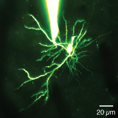
Dendrites, the branch-like projections of neurons, were once thought to be passive wiring in the brain. But now researchers at the Univ. of North Carolina at Chapel Hill have shown that these dendrites do more than relay information from one neuron to the next. They actively process information, multiplying the brain’s computing power.

“Suddenly, it’s as if the processing power of the brain is much greater than we had originally thought,” said Spencer Smith, PhD, an assistant professor in the UNC School of Medicine.

His team’s findings, published October 27 in the journalNature, could change the way scientists think about long-standing scientific models of how neural circuitry functions in the brain, while also helping researchers better understand neurological disorders.

“Imagine you’re reverse engineering a piece of alien technology, and what you thought was simple wiring turns out to be transistors that compute information,” Smith said. “That’s what this finding is like. The implications are exciting to think about.”

Axons are where neurons conventionally generate electrical spikes, but many of the same molecules that support axonal spikes are also present in the dendrites. Previous research using dissected brain tissue had demonstrated that dendrites can use those molecules to generate electrical spikes themselves, but it was unclear whether normal brain activity involved those dendritic spikes. For example, could dendritic spikes be involved in how we see?

The answer, Smith’s team found, is yes. Dendrites effectively act as mini-neural computers, actively processing neuronal input signals themselves.

Directly demonstrating this required a series of intricate experiments that took years and spanned two continents, beginning in senior author Michael Hausser’s lab at University College London, and being completed after Smith and Ikuko Smith, PhD, DVM, set up their own lab at the Univ. of North Carolina. They used patch-clamp electrophysiology to attach a microscopic glass pipette electrode, filled with a physiological solution, to a neuronal dendrite in the brain of a mouse. The idea was to directly “listen” in on the electrical signaling process.

“Attaching the pipette to a dendrite is tremendously technically challenging,” Smith said. “You can’t approach the dendrite from any direction. And you can’t see the dendrite. So you have to do this blind. It’s like fishing if all you can see is the electrical trace of a fish.” And you can’t use bait. “You just go for it and see if you can hit a dendrite,” he said. “Most of the time you can’t.”

But Smith built his own two-photon microscope system to make things easier.

Once the pipette was attached to a dendrite, Smith’s team took electrical recordings from individual dendrites within the brains of anesthetized and awake mice. As the mice viewed visual stimuli on a computer screen, the researchers saw an unusual pattern of electrical signals – bursts of spikes – in the dendrite.

Smith’s team then found that the dendritic spikes occurred selectively, depending on the visual stimulus, indicating that the dendrites processed information about what the animal was seeing.

To provide visual evidence of their finding, Smith’s team filled neurons with calcium dye, which provided an optical readout of spiking. This revealed that dendrites fired spikes while other parts of the neuron did not, meaning that the spikes were the result of local processing within the dendrites.

Study co-author Tiago Branco, PhD, created a biophysical, mathematical model of neurons and found that known mechanisms could support the dendritic spiking recorded electrically, further validating the interpretation of the data.

“All the data pointed to the same conclusion,” Smith said. “The dendrites are not passive integrators of sensory-driven input; they seem to be a computational unit as well.”

His team plans to explore what this newly discovered dendritic role may play in brain circuitry and particularly in conditions like Timothy syndrome, in which the integration of dendritic signals may go awry.

Source:


FDA approves Abbott device for leaking heart valve

The U.S. Food and Drug Administration has approved Abbott Laboratories’ MitraClip medical device, used to stop heart valve leakage in patients deemed unable to endure valve repair through open heart surgery, the company said on Friday.

The MitraClip treats mitral regurgitation, a condition in which the mitral valve of the heart does not close properly, causing blood leakage that can lead to stroke, heart attack or even death.

It has estimated the disorder affects about one in 10 people aged 75 and older.

Those with the condition who are too frail for open heart surgery are typically treated with medicines and have high rates of heart failure and rehospitalizations.

“We think longer term in the U.S., (MitraClip) could be a $500 million product,” said RBC Capital Markets analyst Glenn Novarro. “This approval is sooner than we thought. It’s a pleasant surprise.”

Novarro said the timing of the FDA green light was excellent as it came just ahead of a major U.S. medical meeting for interventional cardiologists where Abbott will be able to showcase the device.

A panel of advisers to the FDA in March voted 5-3 to recommend approval of the implantable heart device. Some panel members questioned whether MitraClip would be effective.

The MitraClip was approved in Europe in 2008 under a system in which medical devices often reach the market several years ahead of the United States.

International sales are running at about $30 million a quarter, with sales growth at about 50 percent over 2012, Abbott said.

U.S. sales are likely to grow slowly at first as the company seeks reimbursement for the device, primarily from the Medicare healthcare program, and as more physicians are trained in its use. The MitraClip is implanted using a minimally invasive procedure in which it is threaded by catheter through a vein into place in the heart to stop the leak.

There are currently 50 centers in the United States that have experience with the device through clinical trials. That number is expected to double over the next year, John Capek, Abbott’s head of medical devices, said in an interview.

There are 20,000 to 30,000 patients in the United States who would likely qualify for MitraClip implantation, Capek said.

Source: