Brain signal that may help in drug de-addiction

braintorm

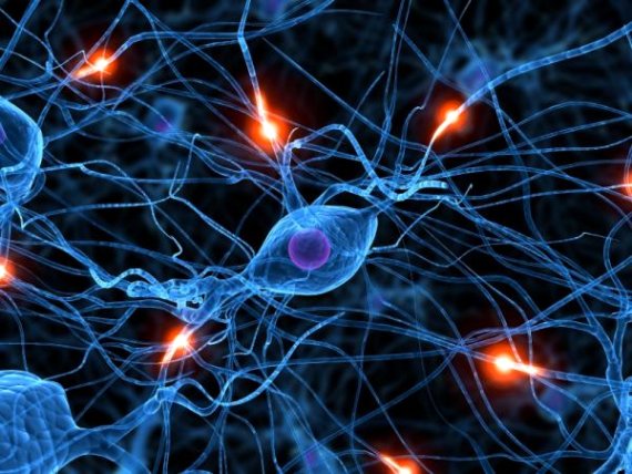
Researchers have discovered a new form of neurotransmission that influences long-lasting memory created by addictive drugs like cocaine and opioids and the craving for them.

Loss of this type of neurotransmission creates changes in brain cells that resemble the changes caused by drug addiction.

The findings suggest that targeting this type of neurotransmission might lead to new therapies for treating drug addiction.

“Molecular therapies for drug addiction are pretty much non-existent,” said Collin Kreple from the University of Iowa in the US.

“I think this finding at least provides the possibility of a new molecular target,” Kreple added.

This neurotransmission involves proteins called acid-sensing ion channels (ASICs), which have previously been shown to promote learning and memory, and which are abundant in a part of the brain involved in drug addiction.
The experiments showed that loss of ASIC signalling increases learned drug-seeking in mice.

When mice learned to associate one side of a chamber with receiving cocaine, animals that lacked the ASIC protein developed an even stronger preference for the “cocaine side” than control mice, suggesting that loss of ASIC had increased addiction behaviour.

The same result was seen for morphine, which has a different mechanism of action than cocaine.

In a second experiment, rats learned to press a lever to self-administer cocaine. Blocking or removing ASIC in the rat brains caused the animals to self-administer more cocaine than control animals.

Conversely, increasing the amount of ASIC by over-expressing the protein seemed to decrease the animals’ craving for cocaine.

The study was published in the journal Nature Neuroscience.

Source: yahoo news


3D brain view may help treat Alzheimer’s, Parkinson’s

3D brain view may help treat Alzheimer's Parkinson's

In a breakthrough that may help in developing drugs for Alzheimer’s and other neurological disorders, researchers have developed a 3D view of an important receptor in the brain.

This receptor allows us to learn and remember, and its dysfunction can result in a wide range of neurological diseases including Alzheimer’s, Parkinson’s, schizophrenia and depression.

The unprecedented view gives scientists new insight into how the receptor – called the NMDA receptor – is structured.

And importantly, the new detailed view gives vital clues for developing drugs to combat neurological diseases and conditions.

“This is the most exciting moment of my career,” said Eric Gouaux, a senior scientist with Oregon Health and Science University in the US.

“The NMDA receptor is one of the most essential, and still sometimes mysterious, receptors in our brain. Now, with this work, we can see it in fascinating detail,” he said.

Receptors facilitate chemical and electrical signals between neurons in the brain allowing them to communicate with each other.

The NMDA (N-methyl-D-aspartate) receptor facilitates neuron communication that is the foundation of memory, learning and thought.

Malfunction of the NMDA receptor occurs when it is increasingly or decreasingly active.

The NMDA receptor makeup includes receptor “subunits” – all of which have distinct properties and act in distinct ways in the brain, sometimes causing neurological problems.

Prior to Gouaux’s study, scientists had only a limited view of how those subtypes were arranged in the NMDA receptor complex and how they interacted to carry out specific functions within the brain and the central nervous system.

Gouaux’s team of scientists created a 3D model of the NMDA receptor through a process called X-ray crystallography.

“This new detailed view will be invaluable as we try to develop drugs that might work on specific subunits and therefore help fight or cure some of these neurological diseases and conditions,” Gouaux said.

“Seeing the structure in more detail can unlock some of its secrets and may help a lot of people,” he added.

The findings were published online in the journal Nature.

Source: Hindustan Times


Indo-Canadian duo develops device to detect drug usage

Indo-Canadian duo develops device to detect drug usage

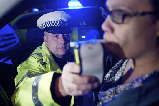
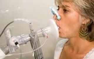
Two Canadians of Indian origin have developed a device that detects marijuana in a person’s breath. Police say it helps to find if a driver is high on the contraband drug.

Vancouver-based Kal Malhi, a former officer of the Royal Canadian Mounted Police (RCMP), the federal police force, has joined hands with Indo-Canadian radiologist Raj Attariwala to develop the device called “breathalyzer” for the police.

A report in the Globe and Mail said Malhi, who worked in the drug investigation branch of the RCMP for four years, believes police badly need a tool that makes it easier to tell if a driver is high.

The breathalyzer is a hand held device that provides accurate reading of marijuana in a person’s breath, says the report. They have applied for the patents of the device and it may take an year before they get it.

“I think it’s more necessary across North America now than it was before, because we’re going through a system when it comes to marijuana, where marijuana is being made readily available across our societies,” Malhi was quoted as saying by the paper.

Colorado and Washington states of teh US have legalised marijuana. So, also the Canadian department of health has allowed medical use of marijuana.

A family doctor can, under certain circumstances, prescribe a dose of up to five grams of marijuana a day (safe dose is 400 mg of Cannabis that contains less than nine percent THC) and should be bought from licensed growers.

After leaving the federal police force in 2011, Malhi reportedly decided to develop the Cannabis Breathalyzer. This test has a major advantage – it can show if a person has smoked marijuana in the past two hours.

According to the report in the Canadian daily, “In saliva and urine tests, marijuana can be detected if it’s been smoked at any point within 72 hours, long after the high has passed.”

“Trying to prove a driver is still high when they’ve gotten behind the wheel,” Malhi said, “can be frustrating for police.”

“It’s largely based on observations you make of the person. And the observations aren’t so obvious for drugged drivers as they are for drunk drivers,” explains Malhi.

Source: healcon


Do autistics struggle with driving?

Do autistics struggle with driving

In the first pilot study asking adults on the autism spectrum about their experiences with driving, researchers at Drexel University found significant differences in self-reported driving behaviors and perceptions of driving ability in comparison to non-autistic adults. As the population of adults with autism continues growing rapidly, the survey provides a first step toward identifying whether this population has unmet needs for educational supports to empower safe driving – a key element of independent functioning in many people’s lives.

“Previous research in my lab has included extensive research in driving capacity with people who have a variety of conditions such as multiple sclerosis or who had experienced traumatic brain injury,” said study co-author Maria Schultheis, PhD, an associate professor of psychology at Drexel. “When we investigate whether and under what circumstances a condition or neurological difference might affect driving ability, as a standard starting point we want to go to individuals and find out from their perspective what problems they are having on the road, in their real-world experience. That question is pivotal to shape and inform the goals of long-term research – and is especially important when we turn to look at a developmental difference like autism, where there has been too little research to establish yet whether widespread driving difficulties exist.”

Only a few previous studies have examined driving ability in individuals with autism, and those studies focused on adolescents and new drivers rather than experienced adult drivers. These studies relied on parent surveys and evaluations of discrete aspects of driving performance. The new Drexel study, published early online this month in the Journal of Autism and Developmental Disorders, used a validated survey that has been extensively used in driving research, and asked adult licensed drivers on the autism spectrum to describe their first-hand, real-world driving experiences.

“We were beginning to see discussion in the research literature that aspects of autism spectrum disorders, such as neurocognitive challenges and social recognition difficulties, could make it likely that members of this population would experience significant challenges with driving,” said the study’s lead author Brian Daly, PhD, an assistant professor of psychology in Drexel’s College of Arts and Sciences. “But that assumption hadn’t been studied in adult drivers, or based on the experiences of the drivers themselves – so these were the questions we explored.”

In this survey, adults with autism spectrum disorders reported earning their drivers’ licenses at a later age, driving less frequently and putting more restrictions on their own driving behaviors (such as avoiding driving on highways or at night), on average compared to non-autistic adults. The respondents with autism spectrum disorders also reported more traffic violations.

Because this pilot study was relatively small and based on self-reports of 78 ASD respondents and 94 non-ASD comparison participants, Schultheis and Daly noted that the differences they found were open to several possible interpretations. Autistic adults may have reported driving less often and restricting their behaviors out of self-awareness of actual difficulties or deficiencies in their driving. These difficulties and/or reduced driving exposure could also explain the higher rate of reported violations.

Alternatively, it is possible that the respondents on the autism spectrum were more honest in their answers, but no worse at driving than everyone else.

“In driving research, it’s well established that people have a positive bias when reporting their own driving skills,” said Schultheis. “Because the study relied on self-reported answers, we can’t rule out whether the respondents with autism were simply being more descriptive and honest about their difficulties than the control group.”

One intriguing finding that Daly and Schultheis noted was that the difficulties adults with autism reported were not clustered in any specific areas, such as problems related to social processing of other drivers’ or pedestrians’ expected behaviors, or difficulties with neurocognitive aspects of driving such as motion perception and reaction time.

“It suggests that the challenges these individuals are facing are more global than specific,” Daly said.

“This is such an important study,” said Paul Shattuck, PhD, an associate professor and director of the research program area in life course outcomes at the A.J. Drexel Autism Institute, who was not involved in conducting the study. “Cognitively-able adults on the autism spectrum face many barriers to full participation in society. Facilitating access to transportation options will increase the capacity for these adults to contribute to their communities.”

Daly and Schultheis are continuing to investigate driving behavior in adults with autism through further research, with funding from the A.J. Drexel Autism Institute, the first autism research center focused on a public health science approach. In the next phase of research, the team is using driving simulation in Schultheis’ lab to objectively capture aspects of actual driving performance in adults on the autism spectrum. Individuals interested in enrolling in these studies should contact schultheis@drexel.edu.

“This is a first step toward identifying, categorizing and quantifying challenges that may exist in this population,” Schultheis said. “What we find will help determine what needs there may be for interventions, from driver education programs to different kinds of training exposures.”

Source: eureka alert


Deep brain stimulation offers hope for OCD patient

Deep brain stimulation offers hope for OCD patient

It seems simple: Walk to the refrigerator and grab a drink.
But Brett Larsen, 37, opens the door gingerly — peeks in — closes it, opens it, closes it and opens it again. This goes on for several minutes.

When he finally gets out a bottle of soda, he places his thumb and index finger on the cap, just so. Twists it open. Twists it closed. Twists it open.

“Just think about any movement that you have during the course of a day — closing a door or flushing the toilet — over and over and over,” said Michele Larsen, Brett’s mother.

“I cannot tell you the number of things we’ve had to replace for being broken because they’ve been used so many times.”

At 12, Larsen was diagnosed with obsessive-compulsive disorder, or OCD. It causes anxiety, which grips him so tightly that his only relief is repetition. It manifests in the smallest of tasks: taking a shower, putting on his shoes, walking through a doorway.

There are days when Larsen cannot leave the house.
“I can only imagine how difficult that is to live with that every single living waking moment of your life,” said Dr. Gerald Maguire, Larsen’s psychiatrist.
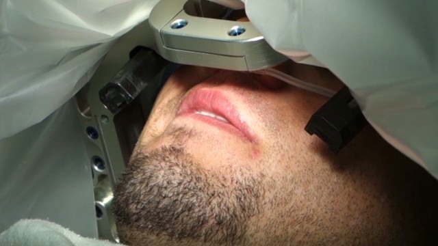
In a last-ditch effort to relieve his symptoms, Larsen decided to undergo deep brain stimulation. Electrodes were implanted in his brain, nestled near the striatum, an area thought to be responsible for deep, primitive emotions such as anxiety and fear.

Brett’s OCD trigger
Brett says his obsessions and compulsions began when he was 10, after his father died.

“I started worrying a lot about my family and loved ones dying or something bad happening to them,” he said. “I just got the thought in my head that if I switch the light off a certain amount of times, maybe I could control it somehow.
“Then I just kept doing it, and it got worse and worse.”

“Being OCD” has become a cultural catchphrase, but for people with the actual disorder, life can feel like a broken record. With OCD, the normal impulse to go back and check if you turned off the stove, or whether you left the lights on, becomes part of a crippling ritual.

The disease hijacked Larsen’s life (he cannot hold down a job and rarely sees friends); his personality (he can be stone-faced, with only glimpses of a slight smile); and his speech (a stuttering-like condition causes his speaking to be halting and labored.)

He spent the past two decades trying everything: multiple medication combinations, cognitive behavioral therapy, cross-country visits to specialists, even hospitalization.

Nothing could quell the anxiety churning inside him. “This is not something that you consider first line for patients because this is invasive,” said Maguire, chair of psychiatry and neuroscience at the University of California Riverside medical school, and part of the team evaluating whether Larsen was a good candidate for deep brain stimulation. “It’s reserved for those patients when the standard therapies, the talk therapies, the medication therapies have failed.”

Deep brain stimulation is an experimental intervention, most commonly used among patients with nervous system disorders such as essential tremor, dystonia or Parkinson’s disease. In rare cases, it has been used for patients with intractable depression and OCD.

The electrodes alter the electrical field around regions of the brain thought to influence disease — in some cases amplifying it, in others dampening it — in hopes of relieving symptoms, said Dr. Frank Hsu, professor and chair of the department of neurosurgery at University of California, Irvine.

Hsu says stimulating the brain has worked with several OCD patients, but that the precise mechanism is not well understood.

The procedure is not innocuous: It involves a small risk of bleeding in the brain, stroke and infection. A battery pack embedded under the skin keeps the electrical current coursing to the brain, but each time the batteries run out, another surgical procedure is required.

‘I feel like laughing’
As doctors navigated Larsen’s brain tissue in the operating room — stimulating different areas to determine where to focus the electrical current — Larsen began to feel his fear fade.

At one point he began beaming, then giggling. It was an uncharacteristic light moment for someone usually gripped by anxiety.

In response to Larsen’s laughter, a staff member in the operating room asked him what he was feeling. Larsen said, “I don’t know why, but I feel happy. I feel like laughing.”

Doctors continued probing his brain for hours, figuring out what areas — and what level of stimulation — might work weeks later, when Larsen would have his device turned on for good.

In the weeks after surgery, the residual swelling in his brain kept those good feelings going. For the first time in years, Larsen and his mother had hope for normalcy.

“I know that Brett has a lot of normal in him, even though this disease eats him up at times,” said Michele Larsen. “There are moments when he’s free enough of anxiety that he can express that. But it’s only moments. It’s not days. It’s not hours. It’s not enough.”

Turning it on
In January, Larsen had his device activated. Almost immediately, he felt a swell of happiness reminiscent of what he had felt in the OR weeks earlier. But that feeling would be fleeting — the process for getting him to an optimal level would take months. Every few weeks doctors increased the electrical current. “Each time I go back it feels better,” Larsen said. “I’m more calm every time they turn it up.”

With time, some of his compulsive behaviors became less pronounced. In May, several weeks after his device was activated, he could put on his shoes with ease. He no longer spun them around in an incessant circle to allay his anxiety.

But other behaviors — such as turning on and shutting off the faucet — continued. Today, things are better, but not completely normal.

Normal, by society’s definition, is not the outcome Larsen should expect, experts say. Patients with an intractable disease who undergo deep brain stimulation should expect to have manageable OCD.

Lately, Larsen feels less trapped by his mind. He is able to make the once interminable trek outside his home within minutes, not hours. He has been to Disneyland with friends twice. He takes long rides along the beach to relax.
In his mind, the future looks bright.

“I feel like I’m getting better every day,” said Larsen, adding that things like going back to school or working now feel within his grasp. “I feel like I’m more able to achieve the things I want to do since I had the surgery.”

Source: cnn


Tiny robotic arm could operate on babies in the womb

surgery in the womb

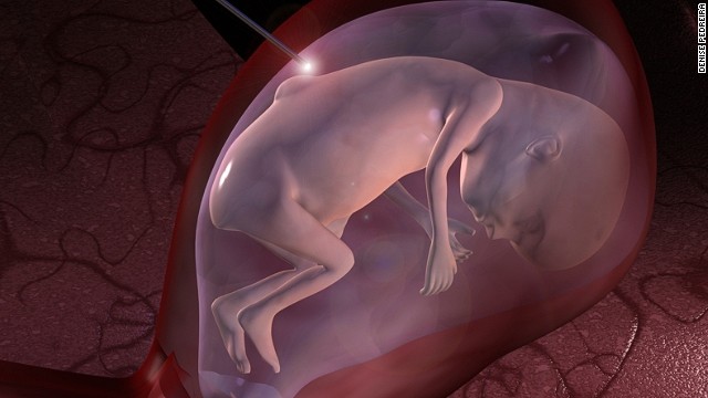
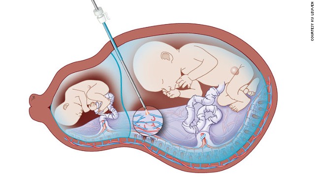
Some birth defects in newborns could one day be a thing of the past due to new robotics technologies being developed to perform surgery on babies in the womb.

Spina bifida is one such disease, affecting approximately 1 in 2,500 newborns worldwide, where a lesion on the back leaves the spinal cord exposed in the womb, leading to severe disabilities, learning difficulties, and sometimes death.
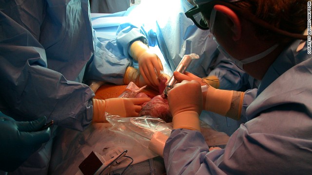
The best option is to perform surgery to correct the problem before the baby is born but the complexities of such a procedure mean this currently only takes place in five countries worldwide. Most countries instead perform surgery after a child is born, but when the majority of damage has been done.

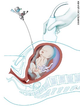
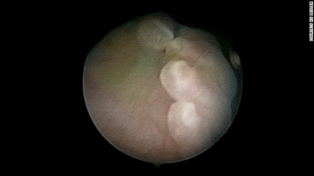
To reduce the risk involved in fetal surgery, scientists at University College London (UCL), and KU Leuven in Belgium are developing a miniscule robotic arm to enter the womb with minimum disruption to mother and baby. The robotics are targeting spina bifida but also lesser known conditions such as twin-twin transfusion syndrome, where blood passes unequally between twins who share a placenta, and fetal lower urinary tract obstruction, where babies are unable to urinate in the womb and their bladders become large and distended.

surgery in the womb1

Surgery on fetuses has been effective in treating some conditions to date, but for spina bifida, the risks to mother and baby mean surgery is currently only performed in a handful of countries, where specialist teams exist.

“Most birth defects can be prevented if we can intervene earlier,” says Professor Sebastien Ourselin, from the UCL Center for Medical Image Computing, who is leading the new research project. “But currently, surgical delivery systems are not available and operating on babies in the womb is reserved for just a handful of the most severe defects as risks are too high.”

Ourselin’s team plans to develop a small three-armed robot, no more than 2 cm wide, to allow more surgeries to take place, as part of a $17 million project funded by the Wellcome Trust and Engineering and Physical Sciences Research Council.

surgery in the womb2

The device will consist of a photoacoustic camera that provides 3D imaging of the fetus in real time, which will help guide two flexible arms to deliver gels or patches to seal the gap in the spine of babies with spina bifida. If successful, the arms will be developed with more dexterity and degrees of freedom to perform surgery themselves and treat further conditions such as congenital heart disease. They may even deliver stem cells as stem cell therapies progress. Once entry into the womb becomes safe, the potential is huge.

In countries where fetal surgery is currently performed, surgeons cut into the mother’s womb before 26 weeks of pregnancy, but there are health risks, side effects to mothers and risks of pre-term labor.

surgery in the womb3

“Where surgery is available in Europe, people are reluctant and fearful of the side-effects,” explains Dr. Jan Deprest, who is leading the work at KU Leuven and has patients declining surgery quite regularly. “Robotic surgery is becoming popular these days and we need to take advantage of that and improve not only the number of patients choosing surgery but also improve the freedom with which we can operate using these flexible probes.”

surgery in the womb4

To have the best effect, surgery must take place before 26 weeks to prevent damage to the exposed spinal cord and the resulting disability. Ourselin wants to go in even earlier.

“We want to go in at 16 weeks to provide the greatest benefit to patients and no one is doing this yet,” concludes Ourselin. “The most important thing is to reduce the invasiveness of the procedure as you want to avoid causing pre-term labor. If we can make this possible, we want to expand treatment to be possible for all diseases which are already present at birth.”

Source: CNN


New breathalyzer test could help detect `deadly` lung cancer

Breath test to detect lung cancer

Researchers have developed a breathalyzer test that could help detect cancer.

The device developed by Prof. Nir Peled of Tel Aviv University’s Sackler Faculty of Medicine, Prof. Hossam Haick (inventor) of the Technion – Israel Institute of Technology, and Prof. Fred Hirsch of the University of Colorado School of Medicine in Denver, is embedded with a “NaNose” nanotech chip to literally “sniff out” cancer tumors.

The study, presented at a recent American Society of Clinical Oncology conference in Chicago, was conducted on 358 patients who were either diagnosed with or at risk for lung cancer. r Peled said lung cancer is a devastating disease, responsible for almost 2,000 deaths in Israel annually – a third of all cancer-related deaths.

He said ” Our new device combines several novel technologies with a new concept – using exhaled breath as a medium of diagnosing cancer.”

Dr Peled said their NaNose was able to detect lung cancer with 90 percent accuracy even when the lung nodule was tiny and hard to sample. It was even able to discriminate between subtypes of cancer, which was unexpected.

“Cancer cells not only have a different and unique smell or signature, you can even discriminate between subtypes and advancement of the disease,” said Dr. Peled. “The bigger the tumor, the more robust the signature.”

The device and subsequent analysis accurately sorted healthy people from people with early-stage lung cancer 85 percent of the time, and healthy people from those with advanced lung cancer 82 percent of the time. The test also accurately distinguished between early and advanced lung cancer 79 percent of the time.

The Boston-based company Alpha Szenszor has licensed the technology and hopes to introduce it to the market within the next few years

Source: ANI


Indian-Origin Scientist Paves Way For Better Epilepsy Treatments

epilepsy

University of Toronto biologists, including one of Indian origin, have discovered proteins to retune imbalances of neurological disorders like autism, epilepsy and various others like schizophrenia and spectrum disorder.

According to Professor Melanie Woodin, the lead investigator of the study, there is a process known as synapses via which neurons in the brain correspond with other neurons, causing neurons either to excite or inhibit other neurons. He further added that any disproportion among the levels of excitation or inhibition may lead to improper brain function.

A crucial complex of protein has been identified that can regulate the proper correspondence of neurons at cellular level. The major proteins are KCC2 which is essential for inhibitory impulse, whereas the receptor for excitatory transmitter glutamate is GluK2, and Neto 2 protein interface with the other two proteins. All three proteins required for synaptic communication is brought together by this complex.

Vivek Madhavan, lead author of the study, along with other researchers conducted experiments on mice brain and found out that all the three proteins directly interact and control each others’ function.

BLUE NATIVE PAGE proved to be the most successful technique of applying a sensitive gel system for determining native protein complexes in neurons.

As such there is no treatment for epilepsy, and the treatments which are available can only curb its effects. Thus the main focus should be on its prevention.

Source: indian nerve


Naloxegol may help treat constipation caused by pain relief medicines

research

A new research has revealed that the drug naloxegol may be able to help patients suffering constipation caused by pain relief medicines.

Globally, approximately 28 million to 35 million, or nearly half, of patients taking opioids for long-term pain develop constipation and they get sub-optimal results from laxatives.

William Chey, the gastroenterologist said that the results showed rapid and sustained improvement in the patients, without compromising their pain management.

Naloxegol is an investigational peripherally-acting mu-opioid receptor antagonist, which has been specifically designed for the treatment of opioid-induced constipation (OIC), a common and often debilitating side effect of prescription medicines used to treat osteoarthritis and chronic back pain.

A New Drug Application (NDA) for naloxegol was accepted by the US Food and Drug Administration on Nov. 19, 2013 and it is also under regulatory review with health agencies in the European Union and Canada.

The study is published online in the New England Journal of Medicine.

Source: Zee news


Erasing (And Restoring) the Brain’s Memories

brain-implant-

In recent years, researchers studying the brain have implanted false memories, caused patients to hallucinate while sleeping, and even tricked the brain into hearing sounds that don’t exist.

Now, a team of neuroscientists has manipulated brain cells to both erase and then restore a memory, a finding that could help with treatment of brain diseases like Alzheimer’s or PTSD.

“Technologically, it’s a huge achievement,” said Steve Ramirez, a graduate student in the department of brain and cognitive sciences at the Massachusetts Institute of Technology who was not part of the study, but has published work on creating memories. “It renders that possibility (of manipulating memory) is no longer science fiction, but something you can tackle experimentally in the lab.”

In a paper published today in the journal Nature, a team at the University of California San Diego created a memory in a rat and then erased it by stimulating the connections, or synapses, between nerve cells at different frequencies.

The first step was to create a memory. They did this by stimulating a group of nerves in the rat’s brain (which equated to the sound of a tone) that had been genetically modified to respond to light, while shocking the animal’s foot at the same time. From the rat’s perspective, the sound of the tone (done by stimulating the nerve cells) was equated with the fear of getting a mild shock.

Then the team weakened the connection between the brain cells, which had the effect of erasing that memory. But in a twist, they were also able to recover the fear-pain memory by strengthening the synaptic connection by stimulating the synapse with a different frequency.

“We can form a memory, and then turn it off and turn it on by selectively turning on synapses,” said Robert Malinow, professor of neurosciences and an author on the paper. “It puts together a number of things we have known and learned to produce this effect. It reinforces that synapses are important and can control memory.”

Malinow says the finding could open the door to manipulating the creation of memories in humans as well. In PTSD, memories of certain traumatic events cause severe anxiety, depression and other problems in patients, while Alzheimer’s disease causes us to lose our memories.

Simulating The Human Brain
A post-doctoral student in Malinow’s lab, Sadegh Nabavi, conducted the experiments and is the lead author on the Nature paper.

One expert, however, cautioned that it’s still too early to take these findings as a road map creating memories in an artificial brain, for example.

“We don’t understand enough about the brain to take those principals and make computers,” said Mark Mayford, a neuroscientist at the Scripps Research Institute in La Jolla, Calif. “Instead, people are trying to take a few principals and put them in standard computer software like learning algorithms.”

Source: discovery news