Scorpion’s venom can make cancer cells ‘glow’

Scorpion’s venom can make cancer cells ‘glow’

In what can alter the course of cancer treatment in the near future, researchers have found a compound that appears to pinpoint all of the malignant cells in a patient’s body. The twist is that the compound’s main ingredient is a molecule that is found in the sting of a deadly scorpion. The compound called chlorotoxin is found in the venom of the death stalker scorpion known as leiurus quinquestriatus. It gives malignant cells a bright fluorescent sheen so surgeons can easily spot them, wired.com reported.

‘A scorpion-venom concoction that makes tumours glow sounded almost too outlandish to be true in the beginning. But with generous donations from individuals, the fluorescent scorpion toxin is now in Phase I clinical trials,’ informed Jim Olson from the renowned Seattle-based Fred Hutchinson Cancer Research Centre that developed the technique, called ‘Tumour Paint’. (Read: Cancer vaccine developed to boost lifespan of patients)

Scorpion venoms are cocktails of numerous individual toxins that attack different targets within a victim’s body. Olson and his team found that chlorotoxin did not attach just to brain tumours — it grabbed onto all sorts of cancers, from those that affect the skin to those that destroy the lungs. In lab experiments, Olson began to inject fluorescent-tipped chlorotoxin into mice — the compound lit up cancer cells that no other technology could identify. In one instance, the chlorotoxin illuminated a clump of just 200 malignant cells that were burrowed deep within a wad of fat. ‘That was the point we learned that the technology was far more sensitive than an MRI,’ Olson was quoted as saying

Source: the health site


Full Moon Night May Reduce Sleep by 20 Minutes

Full Moon Night May Reduce Sleep by 20 Minutes

Next time when your grandmother tells you a folklore as you try to sleep on a full moon night, tell her to cut short as you are going to lose some sleep owing to the effect of lunar cycle on your brain.

Researchers have found that people actually sleep 20 minutes less when the moon is full.

“Participants slept an average of 20 minutes less and had more trouble falling asleep during the full moon phase. However, the greatest impact on REM sleep (during which most dreaming is believed to occur) appeared to be during the new moon,” said Michael Smith from Sahlgrenska Academy at University of Gothenburg in Sweden.

Based on a study of 47 healthy adults aged 18 to 30, the results support an earlier theory that a correlation between sleep and the lunar cycle exists.

“The brain is more susceptible to external disturbances when the moon is full,” Smith added.

A Swiss research study conducted last year showed that the full moon affects sleep.

The findings demonstrated that people average 20 minutes less sleep, take five minutes longer to fall asleep and experience 30 minutes more of REM sleep.

“There may be a built-in biological clock that is affected by the moon, similar to the one that regulates the circadian rhythm,” researchers said.

Re-analysis of the data showed that sensitivity, measured as reactivity of the cerebral cortex in the brain, is greatest during the full moon.

Greater cortical reactivity was found in both women and men whereas only men had more trouble falling asleep and slept less when the moon was full, said the paper that appeared in the journal Current Biology.

Source: ndtv


‘On-off’ switch can help coma patients come to life

on off switch-can-help-coma-patients-come-life

Do you know there is an “on-off” switch in our brain that controls consciousness? Scientists have found one that can help coma patients regain consciousness.

Researchers at George Washington University made an epileptic patient go to sleep by applying an electrical impulse in a specific brain region.

After they stopped the stimulation, the patient came out of coma and had no memory of what had just happened.

“It is like the ignition of a car when turning a key can bring all the other components of the car to life,” Mohamad Koubeissi from George Washington University was quoted as saying.

The effect was produced by stimulating the patient’s claustrum – a thin sheet of neutrons at the lower part of the central brain. Once stimulation of the claustrum stopped, the patient regained consciousness.

Koubeissi found that low-frequency stimulation reduced epileptic seizures in patients by 92 percent without impairing memory.

The discovery of the switch could be very useful in certain areas of medicine, said the study reported by New Scientist.

Source: business standard


Childhood vaccines are safe. Seriously

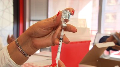
Flu Shots

Children should get vaccinated against preventable and potentially deadly diseases.

That’s what a project that screened more than 20,000 scientific titles and 67 papers on vaccine safety concludes this week. The review appears in the latest edition of the medical journal Pediatrics.

The evidence strongly suggests that side effects from vaccines are incredibly rare, the study authors said. They found no ties between vaccines and the rising number of children with autism, as a small but vocal group of anti-vaccine activists, including actors Jenny McCarthy and Jim Carey, have said.

The review also found no link between vaccines and childhood leukemia, something that was suggested in earlier studies. The researchers found that some vaccines did cause a few adverse effects but it was only for a tiny fraction of the population.

There was evidence that the meningococcal vaccine can lead to anaphylaxis — a severe, whole-body allergic reaction — in children allergic to ingredients in the vaccine. Other studies found the MMR vaccine was linked to seizures.
“Vaccines, like any other medication, aren’t 100% risk free,” said Dr. Ari Brown an Austin, Texas-based pediatrician and author of the popular book “Baby 411,” who was not involved with the study.

“You have a sore arm, redness at the injection site. Those are the things we see commonly. Fortunately the serious adverse effects is extremely rare.” Brown said parents ask her how safe vaccines are all the time. Some patients also ask if they should delay or stagger the vaccinations. She counsels against that practice. She said the younger the child, the more danger these diseases present.

“By delaying the vaccines you’re putting your child at risk,” Brown said. The positive effects of vaccines dramatically outweigh the bad, experts said.

An editorial accompanying the study calls vaccines “one of the most successful public health achievements of the 20th century.” Because of vaccines, many diseases that plagued children for centuries have all but been eliminated.

“There were good reasons that these diseases were targeted for vaccine development since they are so life-threatening,” said Dr. Carrie Byington, vice-chair for research in the University of Utah’s pediatrics department, and the new chair for the American Academy of Pediatrics committee on infectious diseases.

Millions of Americans live longer on average because of the protection vaccines provide. Life expectancy has gone up in the United States by more than 30 years. Infant mortality decreased from 100 deaths per 1000 to 7 between the 1900s and 2000.

A vaccine for smallpox led the Centers for Disease Control and Prevention to declare the disease eradicated in 1978. Prior to a vaccination for diphtheria, it was one of the most common causes of illness and death among children.  Now it is rarely reported in the United States.

What vaccines do children need? Experts: vaccines are necessary
Yet research shows there is still doubt among some medical residents about the effectiveness of vaccinations.

“That is particularly concerning for me,” Byington said. “Young residents may be in the same position as young parents who have trained at a time, or lived at a time, when these diseases were extremely rare, and they may not have ever seen how serious a vaccine-preventable infection can be.”

An increasing number of parents over the years have opted out of getting their children vaccinated. And that may be having a negative impact on the community’s health.

A study found that large clusters of children who had not been vaccinated were close to the large clusters of whooping cough cases in the 2010 California epidemic. While California typically has higher vaccination rates than the rest of the country, that state is dealing with yet another whooping cough epidemic.

This spring also saw an 18-year high number of measles cases in the United States. The largest outbreak was in Ohio where the virus spread quickly among the Amish, who are mostly unvaccinated. This outbreak was a real surprise to health officials who thought that the infectious disease was thought to have been eliminated from the United States in 2000.

The editorial accompanying this latest study suggests doctors, who parents typically trust to tell the truth about medical information, need to use this study to speak with confidence about the importance of vaccinating children.
“Looking at all these mounds of data — there is still no data that show an association that shows vaccine and autism,” said Brown. “I would love it to close this chapter and move on. I don’t think it will. But the more research, the more we learns about autism, the more we can reassure parents that there are no links here.

Source: cnn news


Genes May Be Key to Great Musicians

Genes May Be Key to Great Musicians

Chopin, Vivaldi and Bach may have had natural musical talent, and then some. A new study suggests accomplished musicians are genetically programmed to commit to the long hours of practice needed to become skilled musicians.

The findings add to growing evidence that both nature and nurture help develop expertise, according to the researchers.

“The nature versus nurture debate has raged since the beginning of psychology,” study leader Zach Hambrick, a professor of psychology at Michigan State University, said in a university news release. “This makes it very clear that it’s both. Not only in the sense that both nature and nurture contribute, but that they interact with each other.”

He and his colleagues looked at 850 sets of twins and found that accomplished musicians practiced much more than those who didn’t attain the same level of musical skill, according to the study published online in the June issue of Psychonomic Bulletin & Review.

By comparing identical twins (who share 100 percent of their genes) and fraternal twins (who share 50 percent of their genes), the researchers concluded that an inclination to practice more was driven partly by genetics.

In terms of musical achievement, they also found that genes had a larger effect on those who practiced than on those who didn’t.

The findings challenge the widely held view that a lack of natural ability can be overcome with enough practice and/or training, according to the study authors.

“Contrary to the view that genetic effects go away as you practice more and more, we found that genes become more important in accounting for differences across people in music performance as they practice,” Hambrick said.

Source: web md


Cheap Portable Device to Diagnose TB Faster

Cheap Portable Device to Diagnose TB Faster

Researchers have developed a cheap enzyme-based method that can diagnose tuberculosis (TB) more accurately and faster than available devices.

This inexpensive portable diagnosis system can cut the time it takes to spot TB bacteria from weeks or months to less than half an hour.

Chemist Jianghong Rao of Stanford University and microbiologist Jeffrey Cirillo of Texas A&M University developed a chemical called CDG-3 that glows when it is broken down by an mycobacterium tuberculosis (TB bacteria) enzyme called BlaC.

The researchers found they could detect as few as 10 TB bacteria in a millilitre sample.

They then tested the method on 50 sputum samples from people in Texas.
It correctly identified all the samples that contained M tuberculosis visible under a microscope, and 80 per cent of those in which infections were not visible.

When tested in people without TB, the CDG-3 probe diagnosed them correctly 73 per cent of the time.

Rao and Cirillo are now working to develop a portable, battery-powered device that measures the fluorescence coming from CDG-3 as it is broken down.
The device is expected to hit the market in 2015.

A single test will cost about $5 (Rs 295) and will take less than 30 minutes to deliver a diagnosis, said the paper published in the journal Angewandte Chemie.

A device to cultivate single bacteria species: Millions of microbial species populate the world, but so far only a few have been identified due to the inability of most microbes to grow in the laboratory.

Edgar Goluch, an engineer, and Slava Epstein, a biologist, both from Northeastern University in the US have now developed a device that allows scientists to cultivate a single species of bacteria that can then be studied and identified.

This new device permits just a single bacterial cell to enter an inner chamber containing a food source, to which the only access is a microscopic passageway just slightly narrower than a single cell.

The passageway is so small that the first cell to enter it gets stuck, blocking entry by any other cell or species.

Once inside, this cell proliferate as in previous devices, and when it does it fills up the inner chamber with a pure, single-species sample, since it is isolated from competition from other species.
The researchers demonstrated the device’s ability to separate mixtures of cell types in a laboratory setting.

In one experiment, the researchers separated two different bacterial species whose cells are slightly different sizes — E coli and P aueruginosa.
In a second experiment, they isolated a combination of similarly sized but differently shaped species that commonly show up together in the marine environment — Roseobacter sp and Pscyhoserpens sp.

Finally, they used the device to separate cells of the same species that had been differentially tagged to glow either red or green.
The findings appeared in the journal PLOS ONE.

Source: Oman Observer


Biochemical cascade causes bone marrow inflammation and blood disorders

Biochemical cascade causes bone marrow inflammation and serious blood disorders

Like a line of falling dominos, a cascade of molecular events in the bone marrow produces high levels of inflammation that disrupt normal blood formation and lead to potentially deadly disorders including leukemia, an Indiana University-led research team has reported.

The discovery, published by the journal Cell Stem Cell, points the way to potential new strategies to treat the blood disorders and further illuminates the relationship between inflammation and cancer, said lead investigator Nadia Carlesso, M.D., Ph.D., associate professor of pediatrics at the Indiana University School of Medicine.

Bone marrow includes the cells that produce the body’s red and white blood system cells in a process called hematopoiesis. The marrow also provides a support system and “home” for the blood-producing cells called the hematopoietic micro-environment. The new research demonstrates the importance of the hematopoietic micro-environment in the development of a group of potentially deadly diseases called myeloproliferative disorders.

“It has been known for years that there are links between inflammation and cancer, but these studies have been challenged by the lack of genetic models, especially for blood-based malignancies,” said Dr. Carlesso, a member of the hematologic malignancy and stem cell biology program within the Wells Center for Pediatric Research at IU.

The researchers focused on what happens when there are abnormally low levels of a molecule called Notch, which plays an important role in the process of blood cell production. Using a genetically modified mouse, they found that the loss of Notch function in the microenvironment causes a chain of molecular events that result in excess production of inflammatory factors.

The high levels of inflammation in the bone marrow were associated with the development of a myeloproliferative disorder in the mice. Myeloproliferative diseases in humans can result in several illnesses caused by overproduction of myeloid cells, which are normally are used to fight infections. These diseases can put patients at risk for heart attack or stroke, and frequently progress into acute leukemia and bone marrow failure, which have fatal outcomes. Unfortunately, there are no effective therapies for the majority of myeloproliferative diseases.

When Dr. Carlesso’s team blocked the activity of one of the molecules in this biochemical cascade, the myeloproliferative disorder in the mice was reversed. In addition, elevated levels of the blocked molecule were found in samples from human patients with myeloproliferative disease. These findings suggest that developing drugs that target this inflammatory reaction at different key points could be a promising strategy to limit the development of myeloproliferative disease in humans.

The molecular cascade leading to inflammation was not occurring directly in the bone marrow cells that produce blood cells, but in cells of the bone marrow microenvironment, especially in endothelial cells that line the capillaries — tiny blood vessels — inside the bone marrow. This was a key discovery, Dr. Carlesso said.

“This work indicates that we need to target not only the tumor cells, but also the inflammatory microenvironment that surrounds them and may contribute to their generation,” she said.

“We believe that this combined strategy will be more effective in preventing myeloproliferative disease progression and transformation in acute leukemias.” Dr. Carlesso also noted that the Notch molecule is mostly known as an oncogene — one that can cause cancer — and so is often targeted by therapies for other types of cancer. The new research indicates that clinicians need to be aware of the effects that reducing levels of Notch function could have on the blood development process, she said.

Source: science daily


Researchers regrow corneas, first known tissue grown

Researchers regrow corneas, first known tissue grown from an adult human stem cell

Researchers have identified a way to enhance regrowth of human corneal tissue to restore vision, using a molecule known as ABCB5 that acts as a marker for hard-to-find limbal stem cells. The research is also one of the first known examples of constructing a tissue from an adult-derived human stem cell.

Boston researchers have identified a way to enhance regrowth of human corneal tissue to restore vision, using a molecule known as ABCB5 that acts as a marker for hard-to-find limbal stem cells. This work, a collaboration between the Massachusetts Eye and Ear/Schepens Eye Research Institute (Mass. Eye and Ear), Boston Children’s Hospital, Brigham and Women’s Hospital and the VA Boston Healthcare System, provides promise to burn victims, victims of chemical injury and others with damaging eye diseases. The research, published this week in Nature, is also one of the first known examples of constructing a tissue from an adult-derived human stem cell.

Limbal stem cells reside in the eye’s basal limbal epithelium, or limbus, and help maintain and regenerate corneal tissue. Their loss due to injury or disease is one of the leading causes of blindness. In the past, tissue or cell transplants have been used to help the cornea regenerate, but it was unknown whether there were actual limbal stem cells in the grafts, or how many, and the outcomes were not consistent.

In this study, researchers were able to use antibodies detecting ABCB5 to zero in on the stem cells in tissue from deceased human donors and use them to regrow anatomically correct, fully functional human corneas in mice.
“Limbal stem cells are very rare, and successful transplants are dependent on these rare cells,” says Bruce Ksander, Ph.D., of Mass. Eye and Ear, co-lead author on the study with post-doctoral fellow Paraskevi Kolovou, M.D. “This finding will now make it much easier to restore the corneal surface. It’s a very good example of basic research moving quickly to a translational application.”

ABCB5 was originally discovered in the lab of Markus Frank, M.D., of Boston Children’s Hospital, and Natasha Frank, M.D., of the VA Boston Healthcare System and Brigham and Women’s Hospital, co-senior investigators on the study, as being produced in tissue precursor cells in human skin and intestine. In the new work, using a mouse model developed by the Frank lab, they found that ABCB5 also occurs in limbal stem cells and is required for their maintenance and survival, and for corneal development and repair. Mice lacking a functional ABCB5 gene lost their populations of limbal stem cells, and their corneas healed poorly after injury.

“ABCB5 allows limbal stem cells to survive, protecting them from apoptosis [programmed cell death],” says Markus Frank. “The mouse model allowed us for the first time to understand the role of ABCB5 in normal development, and should be very important to the stem cell field in general.” according to Natasha Frank.

Markus Frank is working with biopharmaceutical industry to develop a clinical-grade ABCB5 antibody that would meet U.S. regulatory approvals. “A single lab cannot do a study like this,” says Natasha Frank, also affiliated with the Harvard Stem Cell Institute. “It integrates genetics, knockout mice, antibodies, transplantation — a lot of technical expertise that we were lucky came together in a very nice way.”

Source: science daily


Organ transplants: ‘Supercooling’ keeps organs fresh

‘Supercooling’ keeps organs fresh

A new technique can preserve organs for days before transplanting them, US researchers claim. “Supercooling” combines chilling the organ and pumping nutrients and oxygen through its blood vessels.

Tests on animals, reported in the journal Nature Medicine, showed supercooled livers remained viable for three days, compared with less than 24 hours using current technology.

If it works on human organs, it has the potential to transform organ donation. As soon as an organ is removed from the body, the individual cells it is made from begin to die.

Cooling helps slow the process as it reduces the metabolic rate of the cells. Meanwhile, surgeons in the UK carried out the first “warm liver” transplant in March 2013 which used an organ kept at body temperature in a machine.

The technique being reported first hooks the organ up to a machine which perfuses the organ with nutrients. It is then cooled to minus 6C.

Supercool

In experiments on rat livers, the organs could be preserved for three days. One of the researchers, Dr Korkut Uygun, from the Harvard Medical School, told the BBC the technique could lead to donated organs being shared around the world.

“That would lead to better donor matching, which would reduce-long term organ rejection and complications, which is one of the major issues in organ transplant,” he said.

He also argued that organs which are normally rejected, as they would not survive to the transplant table, might be suitable if they were preserved by supercooling.

“That could basically eliminate waiting for a organ, but that is hugely optimistic,” Dr Uygun said. Further experiments are now needed to see if the technology can be scaled up from preserving a 10g (0.35oz) rat liver to a 1.5kg (3.3lb) human liver.

The researchers believe the technology could work on other organs as well.

Dr Rosemarie Hunziker, from the US National Institute of Biomedical Imaging and Bioengineering, said: “It is exciting to see such an achievement in small animals by recombining and optimizing existing technology.

“The longer we are able to store donated organs, the better the chance the patient will find the best match possible, with both doctors and patients fully prepared for surgery.

“This is a critically important step in advancing the practice of organ storage for transplantation.”

Source: updated news


Decline of hearing ability: Indian, US experts find gene role

Listening

In a path-breaking research which may have implications for those suffering from a decline of their cognitive and hearing abilities, Indian and American experts have established the role of a specific gene in triggering such conditions.

Experts of Sir Ganga Ram Hospital and University of Louisville School of Medicine stated that the MMP-9 gene plays a major role in causing decline of cognitive and hearing functions and removal of the said gene decreases
Hyperhomocysteinemia-induced cognitive and hearing dysfunctions.

Hyperhomocysteinaemia (HHcy) is a medical condition arising due to an abnormally high level of homocysteine in the blood, experts said.

“There is a role of MMP-9 in decline of cognitive and hearing functions. The ablation of MMP-9 decreases Hyperhomocysteinemia-induced cognition and hearing

dysfunction. This research was carried out on mice but has large implication for humans,” said Dr Seema Bhargava, lead author of the research and Senior Consultant, Department of Biochemistry, Sir Ganga Ram Hospital.

MMP-9 gene is a matrix metallopeptidase which helps in wound healing, cell migration, learning, memory and various other functions.

Currently, 45 per cent of adults in India between 45-92 years of age suffer from hearing impairment. Deficiency of Vitamin B-12 and folate (another form of vitamin) and high homocysteine levels have also been associated with impaired
hearing in women.

“It is important to identify individuals at risk for HHcy (e.g. elderly people)… To reduce homocysteine levels, adequate vitamin supplements should be given. However, if HHcy is already present, vitamins will take several months to reduce the concentration of homocysteine.

“Our study has advocated the role of MMP-9 inhibitors by pharmaceutical companies as a therapeutic option,” Bhargava said.
The research was published in the May edition of Journal of Molecular Biology Reports.

Source: Zee news