Honey not a contender in the fight against superbugs

If you haven’t heard about the threat “superbugs” (bacteria that are resistant to antibiotics) pose to our health, it’s likely you haven’t been conscious or on the planet for a couple of years.

There are no new drugs in the pipeline to fix the superbug problem. And when times are tough, and we’re faced with life-threatening infections, we often turn to alternative, sometimes unproven methods.

Research recently published in The Lancet looked at whether applying medical grade honey to wound sites in patients undergoing dialysis showed advantages over standard antibiotic use.

We are susceptible to bacterial infection whenever our skin is punctured, which happens quite a lot during hospital treatment. The researchers worked with dialysis patients because having a catheter inserted is a regular procedure for the hundreds of thousands of kidney dialysis patients.

They found that using honey showed no advantages over standard antibiotic use, and was, in fact, worse for diabetic dialysis patients.

Getting something like honey to the market as a food product is pretty straightforward, but for medicines the bar is set higher.

The drug approval process

There are many different ways that new medicines are approved for public use.

Drugs made by pharmaceutical companies, for instance, go through many years of expensive, highly-controlled clinical trials comparing the effects of the new drug against a placebo control. And later, they are compared against competitor compounds already on the market.

But alternative, or “natural” medicines can be put straight to market provided they don’t do any harm and their makers don’t make outrageous claims about their health effects.

One such natural alternative is bee honey, which has long been known to have antibacterial activity. A commercial version of “antibiotic” honey, Medihoney, has actually been clinically evaluated for the treatment of ulcers.

Honey is cheap and widely available, and while it cannot be used for systemic infections (for pneumonia, for instance, or bacteremia), it has been used for open wounds. It can kill many types of bacteria and is thought also to provide a barrier to moisture.

The bake off

The Lancet paper looked closely at how a naturally-derived honey preparation compares against a clinically-approved antibiotic – a drug called Bactroban that’s used to kill bacteria found in and around wounds.

The main culprit, Staph aureus, lives on our skin and can cause infection around catheters used during treatment of dialysis-related infections.

A total of 371 trial participants undergoing dialysis received either standard antibiotic therapy to prevent infection, or a daily application of medical grade honey, to the site of catheter insertion.

The study found no significant differences in infection rates or deaths from infection between most people given the antibiotic or people given honey.

But for people with diabetes, which is often associated with kidney disease, the honey actually increased the risk of infection. This is important because diabetes is the leading cause of kidney failure, so any therapy should work with normal and diabetic kidney dialysis patients.

The reasons for the diabetes-kidney failure link are not completely understood, but diabetics often have both high blood glucose levels and high blood pressure, both of which can cause kidney damage.

There’s also a hormone system called the renin-angiotensin system that regulates blood pressure and fluid balance involving the kidney, which is unbalanced in diabetics.

While honey therapy was worse than antibiotics for diabetics, the authors found the most important factor for preventing infection in all patients was how well the catheter was inserted and fixed. There was no placebo control possible in the trial (where no treatment is given) as these infections are sometimes fatal.

We can conclude that honey therapy instead of antibiotics at least does no harm for many dialysis patients, but also that it’s not good for diabetics. And because diabetes and kidney disease are commonly linked and 9% of the patients treated with either therapy still died, the bottom line is still that we need better therapies for bacterial infection – whether natural or man-made.

Source:


Study finds reservoir of hidden HIV bigger than once thought

Over the past decade, scientists have made incredible strides in the field of HIV research – leading to the development of numerous medications that can effectively manage the disease and provide patients with a near normal life expectancy.

But a cure for the human immunodeficiency virus has still eluded scientists, and now a new report from Howard Hughes Medical Institute (HHMI) has revealed that completely eradicating the virus may be much more difficult than previously thought.

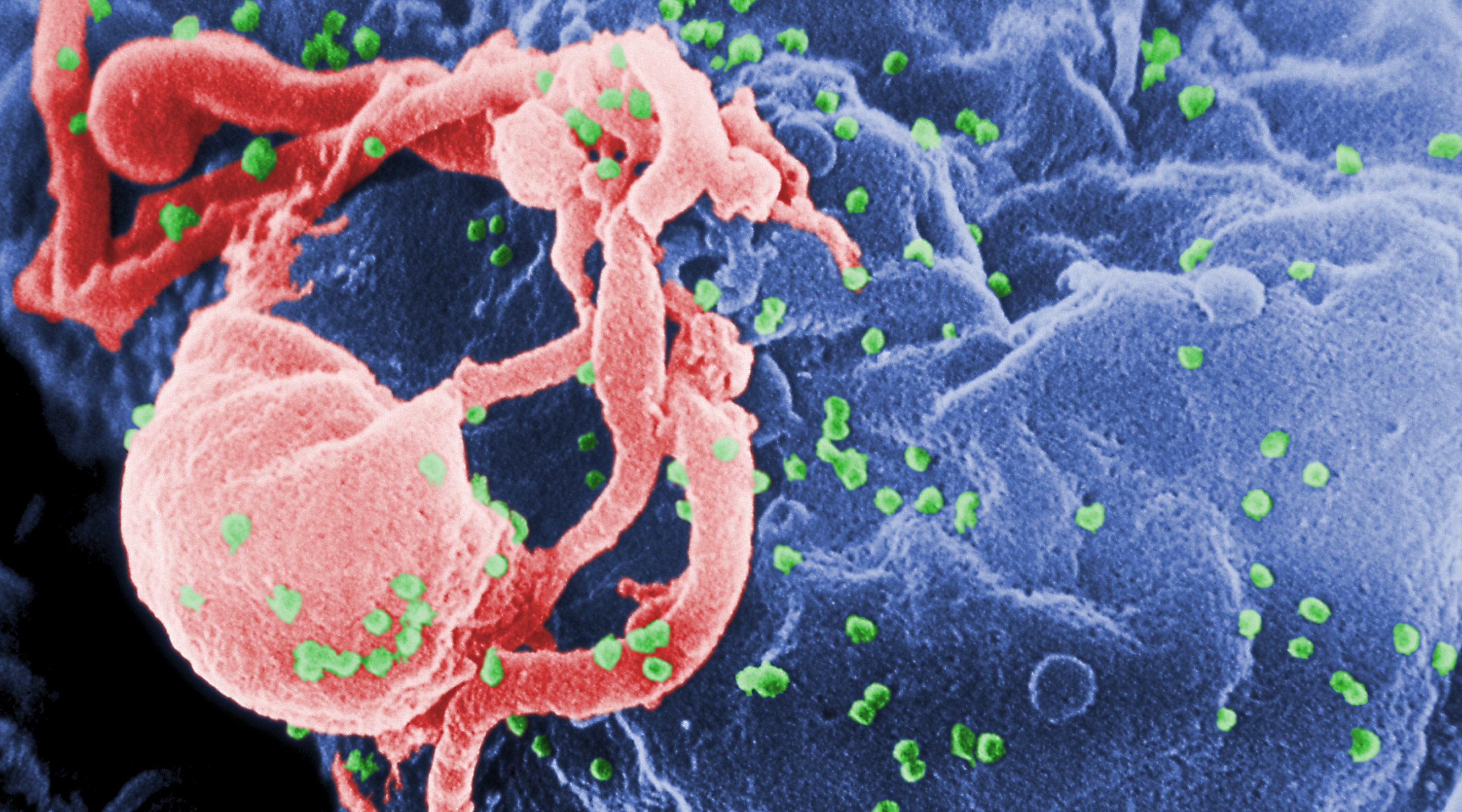
In a new study published in the journal Cell, researchers have discovered that the reservoir of latent or inactive HIV that silently lingers in a patient’s body is much larger than scientists believed.  In fact, this pool of viruses – known as HIV proviruses – may actually be 60-fold greater than previous estimates.

According to researchers, these findings are a major setback in the fight to cure HIV.

“We’re working very hard on developing better ways to assess the size of this reservoir,” lead author Dr. Robert Siliciano, an HHMI investigator at Johns Hopkins University, told FoxNews.com.  “But I think there’s a lot more we really need to understand before we do a lot more clinical trials on (HIV cures).”

In order to effectively cure someone of HIV, the entirety of the virus must be eliminated from the body.  This has been difficult to accomplish, since HIV exists in two states – an active version that is constantly replicating and destroying the immune system and an inactive version that hides in the body’s memory T cells and remains dormant.

When memory T cells respond to the virus and try to eradicate it from the body, they inadvertently become hosts for HIV. The virus “infects” some of the responding T cells by physically inserting itself into the cells’ DNA.  Then, when the T cells go back to a resting state, the HIV is “turned off” and silently hides in its host cell.

Current antiretroviral medications for the treatment of HIV work by targeting the actively replicating form of HIV – but not this inactive form.

“In resting state…the virus is not actively replicating, so the drugs don’t affect it, and the immune system can’t see it, because no viral proteins are being made,” Siliciano explained.  “That’s why you can’t cure the infection, because as soon as the patient stops treatment, some of these memory T cells get activated every day, and they start to replicate, and the virus takes off again.”

Finding the true size of HIV

Previously, researchers would estimate the amount of these latent proviruses by removing T cells from an HIV positive patient and activating them in test tubes.  This approach was thought to force all of the functioning inactive proviruses hiding in the T cells to “turn back on.” Using this technique, approximately one HIV provirus was reactivated out of every million T cells.

But Siliciano and his colleague Ya-Chi Ho, an HHMI international student research fellow in Siliciano’s lab, believed this method did not provide the whole picture, since some of the proviruses remained inactive or latent in the T cells even in response to activation.

So the researchers developed a technique to further analyze the composition of the viral reservoir. In order to do this, they first activated the T cells but then focused on the non-induced proviruses.  Using a genetic technique called polymerase chain reaction (PCR), the researchers were able to study the genomes of the proviruses that had failed to turn on, which remained in 300 out of ever million T cells.

While 88 percent of the non-induced proviruses contained some obvious mutation or defect in their DNA preventing them from replicating, approximately 12 percent had fully intact genomes, meaning they were entirely capable of being reactivated.  And after a second and third attempt at stimulating the T cells, Siliciano and Ho found that these proviruses did, in fact, become active.

“If you take the T cells that did not produce virus, and simply reactivate them a second time, some start to produce virus,” Siliciano said. “…So the total number of cells you have to worry about is any cell that could turn on the virus…Maybe there’s some random process governing this, but those are the things that are not understood.”

These findings mean that scientists have many more proviruses to kill in order to fully cure an individual of HIV.  However, Siliciano hopes his research will dissuade patients from entering into clinical trials testing a “shock and kill” approach to curing the virus, which involves activating many of the body’s T cells to force the proviruses to activate.  Since the size of the provirus reservoir is so large, Siliciano said this technique could lead to major damage in the body.

“The current approach to curing patients is to try and turn on all of the latent virus and then get the cells to die,” Siliciano said. “You can’t do that in a patient because it would cause too many side effects.  Having all your T cells turn on at the same time would lead to major toxicity.”

The HHMI study was released just as recent tests have found that a baby born with HIV in Mississippi is still in remission 18 months after being aggressively treated, potentially indicating that the child is cured.  While Siliciano said this is great news and extremely important for HIV research, there is still much more to learn about the virus before a true cure can be developed.

“In the (Mississippi child’s) case, there are no memory T cells at the time of birth, and that’s why the infection is curable if you treat on the first day of life,” Siliciano said. “So for the vast majority of patients to cure them, we’ll have to deal with this reservoir. These cases of cures are exciting and have given people the feeling that ‘Yes, maybe this is something we can do.’  But it’s going to be a long and difficult struggle.”

Source:


Scientists discover protein that may help prevent cataract

The human eye lens consists of a highly concentrated mix of several proteins. Protective proteins prevent these proteins from aggregating and clumping. If this protective function fails, the lens blurs and the patient develops cataracts. Two research groups at the Department of Chemistry of the Technische Universitaet Muenchen (TUM) have succeeded in explaining the molecular architecture of this kind of protective protein.

Their findings, are published online in the current early edition of PNAS (Proceedings of the National Academy of Sciences), shed new light on the work of these proteins and may be able to help in the development of new treatments.

Cells have a variety of protein complexes that manage vital tasks. The functions of these “molecular machines” depend largely on their three-dimensional structure. In the first instance, proteins are long chains of amino acids, like a long piece of woolen thread. So-called chaperones help them to fold in the desired three-dimensional form after their production. If this folding process fails, the protein thread becomes an inextricable, useless tangle.

Small heat shock proteins (sHsps) are a particularly important group of chaperones. They prevent the clumping of proteins under stress conditions. αB-crystallin and the related sHsp αA-crystallin are the main representatives of the sHsps found in humans. Whereas αA-crystallin mainly occurs in the eye lens, αB-crystallin is also very common in the brain and in the heart and muscle tissue. In the eye lens, they counteract diseases like cataracts. Malfunctions of the αB-crystallin in tissue cells can give rise to cancer and neurological defects, including Alzheimer’s disease.

Many research groups have focused their work on the α-crystallins due to their medical relevance. Despite intensive efforts, up to now, none of them have managed to determine the molecular architecture of these proteins. However, TUM biochemists have now succeeded in producing αA-crystallins and αB-crystallins recombinantly in bacteria and in obtaining uniform, clearly-structured complexes. A detailed structural analysis of these proteins was carried out in cooperation with the Chemistry Department’s Center of Electron Microscopy. The research groups were able to show for the first time here that, contrary to previous suppositions, αB-crystallin forms a defined globular structure comprising 24 subunits, which are reminiscent of a perforated soccer ball.

Thanks to the identification of the three-dimensional structure of αB-crystallin, which is currently being further refined, the basis has now been established for comparing healthy and disease-promoting mutants and, based on this, for clarifying the way they function. The scientists hope that this will lead to the discovery of new treatments.

Source:


World’s first tissue-engineered trachea implant – success 5 years later

Five years after the world’s first tissue-engineered trachea implant, researchers are declaring the procedure is still a success, with the patient reporting good quality of life and minimal complications, Medical Xpress reported.

The patient, a then-30-year-old mother of two from Colombia, received the implant in 2008 after her own trachea collapsed due to complications from tuberculosis. The implant was created using cells from the trachea of a human donor combined with cartilage cells derived from the patient’s own stem cells and epithelial cells taken from the patient’s trachea.

Ten days after the groundbreaking procedure, the patient was discharged from the hospital. After four months, the transplant appeared to be successful, and the patient did not require any immunosuppressive drugs.

In a study published in The Lancet, researchers report that five years after the initial transplant, the recipient is still doing well and enjoying a normal life. Testing has also revealed that the patient has good lung function and isn’t demonstrating any immunological complications, according to Medical Xpress.

Although scarring on the graft required a stent to be inserted six months after the procedure, the patient is no longer experiencing any symptoms of this minor complication, researchers reported.

“These results confirm what we – and many patients– hoped at the time of the original operation: that tissue engineered transplants are safe and effective in the long term,” said Paolo Macchiarini from the Karolinska Institutet in Stockholm, Sweden, who operated on the patient.

Macchiarini noted that he and his fellow researchers hope to improve upon the procedure in the future to avoid complications like the minor scarring seen in the patient. They also hope to continue with clinical trials in order to further demonstrate that this type of procedure can be integrated into routine practice.

According to MedPage Today, several other successful trachea implants have been completed since 2008, including one on a 2-year-old South Korean girl born without a windpipe and another on a 36-year-old cancer patient.

Source:


Study Shows How Melanoma can Become Drug Resistant

A process involving the phenotypes of tumor cells could change the appearance of melanoma tumors, say researchers. Identifying the phenotype patients exhibit may help determine which patients are more likely to benefit from existing medications while also providing an opportunity to create new targeted therapies.

Senior correspond author Ashani Weeraratna, Ph.D., Assistant Professor in the Tumor Microenvironment and Metastasis Program of Wistar’s NCI-designated Cancer Center, and her team focus on Wnt5A, a Wnt signaling molecule that has been found in increased levels in metastatic melanomas.

In order for Wnt5A to promote the phenotype switch from early in the tumor’s formation to the time it becomes metastatic, the tyrosine kinase receptor ROR2 is required. When ROR2 is not present, Wnt5A is unable to promote tumor metastasis.

The only other member of the family that has been identified is ROR1, and this research was done to determine what role ROR1 might play in the progression of melanoma.

The researchers were able to determine that ROR1 inhibited the invasion of melanoma cells, and this receptor was targeted for degradation by Wnt5A and ROR2.

When ROR1 was silenced, the researchers observed that there was an increased rate of invasion of melanoma cells both in vitro and in vivo. The researchers also found that hypoxia – areas of low oxygen supply in the tumor – is able to induce a switch from ROR1 to ROR2 and results in an increase in levels of Wnt5A, suggesting the switch from a non-invasive ROR1-positive phenotype to an invasive ROR2-positive phenotype occurs when the tumor is exposed to hypoxic conditions.

The researchers also found that a protein HIF1a is required to increase the Wnt5A expressed. When HIF1a was removed, ROR2 was decreased, indicating that the up regulation of ROR2 via HIF1a requires Wnt5A.

The findings have been published online in the journal Cancer Discovery.

Read more: http://www.medindia.net


Study shows how Staph toxin disarms the immune system

Researchers at NYU Langone Medical Center have discovered a new mechanism by which the deadly Staphylococcus aureus bacteria attack and kill off immune cells. Findings, published in the journal Cell Host & Microbe, explain a critical survival tactic of pathogen that cause more skin and heart infections than any other microbe and kills more than 100,000 Americans every year.

“What we’ve found is that Staph unleashes a multi-purpose toxin capable of killing different types of immune cells by selectively binding to surface receptors,” says Victor J. Torres, PhD, assistant professor of microbiology, and senior author of the study. “Staph has evolved the clever ability to target the immune system at different stages.”

Scientists have long known that Staph releases an arsenal of toxins to puncture immune cells and clear the way for infection. But only recently have they begun to understand exactly how these toxins work. Earlier this year, Dr. Torres and his team published a paper in Nature explaining how one of those toxins, a protein called LukED, fatally lyses T-cells, macrophages and dendritic cells, all types of white blood cells that help fight off infection. The LukED toxin, they showed, binds to a surface receptor called CCR5 (the same one exploited by HIV). “It attaches to the surface receptor and then triggers pore formation,” says Dr. Torres. But their discovery failed to explain how the bacterial toxin kills other types of white blood cells, such as neutrophils, that lack the CCR5 receptor.

Their most recent work solves this puzzle, showing for the first time how receptors on neutrophils (a common type of white blood cell) also enable binding of the LukED toxin. The researchers found that LukED latches onto surface receptors called CXCR1 and CXCR2, creating the same deadly pores that it does when it latches onto CCR5 receptors. “The mechanism is the same,” says Dr. Torres. “The strategy makes Staph deadlier in mice.”

Neutrophils are the first responders. Upon infection, they race through the bloodstream to kill off the invading pathogen. “They’re like the marines of the immune system,” Dr. Torres says. T-cells, macrophages and dendritic cells rush in later, mounting a secondary attack to help the body clear the pathogen and remember it in the future. “Killing off the first responders completely disarms the immune system,” Dr. Torres says.

LukED is just one piece of the puzzle, and more research is needed to understand other Staph toxins and how they work together to make the microbe deadlier. However, these recent insights hold promise for new medications that target LukED. Better treatments against Staph are desperately needed. In 2005, the Centers for Disease Control and Prevention estimated that more than half of the 478,000 people hospitalized for staph infections were resistant to methicillin, one of the most potent antibiotics available.

One therapeutic strategy is to block CCR5 receptors and spare the secondary immune response. “We know we can block CCR5 receptors without crippling the rest of the immune system. Some people lack CCR5 and they are perfectly healthy and immune to HIV as well,” Dr. Torres says. “But just blocking CCR5 isn’t enough.” Drugs are available to block CXCR1 and CXCR2 receptors, but those will impair neutrophil recruitment and function. “The lesson is to target the toxin itself and prevent it from attaching to any receptors,” Dr. Torres adds. “We have to think globally.”

Source:


Neanderthals used toothpicks to alleviate the pain of diseases related to teeth

An IPHES (Institut Català de Paleoecologia Humana I Evolució Social) research done in collaboration with UAB, documents the oldest case of palliative treatment of periodontal disease. Published in PLOS ONE, is another step to characterize the Neanderthals as a species with a wide range of adaptations to the environment and, even, resources in medicine. 

Removing food scraps trapped between the teeth one of the most common functions of using toothpicks, thus contributing to our oral hygiene. This habit is documented in the genus Homo, as early as Homo habilis, a species that lived between 1.9 and 1.6 million years ago. A new research based on the Cova Foradà Neanderthal fossil shows that this hominid also used toothpicks to mitigate pain caused by oral diseases such as inflammation of the gums (periodontal disease). It is the oldest documented case of palliative treatment of dental disease done with this tool.

The research, done by  Marina Lozano, Carlos Lorenzo and Gala Gomez of the IPHES (Institut Català de Paleoecologia Humana I Evolució Social), in collaboration with Maria Eulalia Subirà, Biological Anthropology professor and researcher at the UAB, and José Aparicio of the Diputació Provincial de València, is based on toothpicking marks on the Neanderthal teeth related to periodontal disease.

The chronology of the fossil is not clear, but the fossil remains were associated with a Neanderthal Mousterian lithic industry (about 150,000 to 50,000 years). The research showed that the remains had maxillary porosity, characteristic of periodontal disease and alveolar bone loss (where the teeth are inserted), with a bone mass reduction of four to eight millimeters exposing the roots of the teeth, usually inside the alveoli.

Resrachers believe that this individual attempted to alleviate the discomfort caused by periodontal disease. This disease usually causes bloody and inflamed gums, so the systematic use of toothpicks could mitigate sore gums.

The examples of grooves caused by toothpicking are numerous between Neanderthals and usually are not associated with any dental disease. However, in the case of Cova Foradà the toothpick was not only used as a primitive method of dental hygiene, but it is associated with a dental disease and with the clear intention to alleviate the pain, and that makes it unique.

This means that we have one of the first examples of palliative treatment with toothpicks, the oldest documented. Therefore, this study is a step to characterize the Neanderthals as a species with a wide range of adaptations to their environment and wide resources even in the field of palliative medicine.

Source: http://www.uab.es


Family’s Untrained Pet Dog ‘Predicts’ Toddler’s Epileptic Fits

Animals have aided the blind for quite some time now. More recently, they have been trained to help calm children with autism and detect health conditions like cancer and diabetic episodes. However, there’s little evidence to indicate that dogs can actually detect epileptic seizures. But you couldn’t tell an Irish family that. They say that Charlie, their Great Dane, can detect their daughter’s epileptic fits before they happen.

Three-year-old Brianna Scanlan, whose episodes were detected at just three months of age, has had epilepsy since birth. Her seizures can range anywhere from a trance-like state where she simply stares off into space to full-on violent convulsions that put her at risk for bodily harm.

“If you see a child having a seizure, it’s pretty horrific, it’s frightening, it’s terrible, it’s gut-wrenching,” Brianna’s mother, Arabella Scanlan told BBC News.

But with Charlie around, the family feels just a little safer.

According to Arabella, Charlie is not a trained “seizure alert dog.” In fact, he’s just a normal family pet. But some time ago, the family started to notice that Charlie would become agitated and walk in circles around Brianna, just minutes before a seizure would happen.

“Charlie would know about 15 to 20 minutes before she’s going into a seizure,” Arabella said. “He’ll get ever so panicky and giddy, almost as if you’d think ‘this stupid dog is going to knock her over.’”

In fact, the family initially thought they would have to re-home Charlie because of his agitation. He is, after all, a Great Dane.

“He’s a big boy – it isn’t like he’s agile. When Charlie turns the whole room turns with him,” Arabella said.

But as the family watched Charlie, they started to notice a pattern in his behavior. His agitation seemed to precede Brianna’s epileptic fits.

“We kept an eye on this and, sure enough, I went into the yard one day and she (Brianna) was buckled over to the side, on top of him (Charlie). She was actually having a seizure,” Arabella said. “She was leaning against the wall, bent over him and he just looked at me as if to say ‘I don’t know what to do.’ But he stayed with her, he didn’t move.”

Since then, the family says that Charlie rarely ever leaves Brianna’s side. If she goes into a seizure, he’ll gently pin her against the wall or another surface to keep her from falling over. He’ll then guard her and watch over her until help arrives. And, according to Arabella, he hasn’t once knocked her over.

“I actually don’t know the psychology behind it but, no shadow of a doubt; people are mesmerized when they see him in action. It would actually melt you hear to see them together.”

Unfortunately, there isn’t much in the way of science to back up this family’s amazing story. Though dogs have been found to “sense” a number of health issues – dysregulation in individuals with autism, blood sugar spikes and drops in diabetic patients, cancers and more – there’s only been a few studies done on dogs and epilepsy.

One preliminary study, published in Seizure, European Journal of Epilepsy, suggested that “some dogs have innate ability to alert and/or respond to seizures.” It added that the success of this ability was found to rely heavily “on the handler’s awareness and response to the dog’s alerting behavior.”

Yet, the lack of scientific information regarding a dog’s ability to sense seizures doesn’t stop the Sheffield-based charity, Support Dogs, from training “seizure alert dogs.” According to the training facility, their trained dogs are able to detect seizures anywhere from 10 to 55 minutes before it occurs.

Dr. Claire Guest, a Medical Detection Dogs chief executive, says that she’s seen other trained dogs in action. In fact, she even had a cancer dog “warn her” during training, and that dog ended up being right; a little while later, she was diagnosed with first stage breast cancer. But Guest says she’s never really seen a dog predict an epileptic fit. However, she did state that it’s not impossible.

Dogs that are highly expressive and attentive to humans tend to show a general concern and desire to protect their owners from harm, and it could be that some dogs could predict seizures through smell, but it’s also likely that they’re picking up on visual signs, she said.

Source:


Sustained protection against cholera in humans up to five years.

The study showed the vaccine had a protective efficacy of 65 percent over a five-year period.

The landmark study was collaboration between scientists from the International Vaccine Institute (IVI) an international organization based in Seoul, and the National Institute of Cholera and Enteric Diseases, (NICED), an institute under the Indian Council of Medical Research (ICMR) of India’s Ministry of Health and Family Welfare.

Cholera is a potentially deadly infectious disease that causes profuse, dehydrating diarrhea in children and adults.

The oral cholera vaccine (OCV) contains strains of killed cholera bacteria that have been previously shown to be safe in humans and is administered through a two-dose regimen.

The vaccine was specifically developed for use in developing countries through a public-private partnership led by IVI with support from the Republic of Korea, Sweden, and the Bill and Melinda Gates Foundation.

The partnership involved Shantha Biotechnics (part of the Sanofi group) based in Hyderabad, India; VaBiotech, a state-owned vaccine manufacturer located in Hanoi, Vietnam; and the University of Gothenburg in Sweden.

The vaccine, which is produced by Shantha Biotechnics in India and licensed as Shanchol, was prequalified by theWorld Health Organization (WHO) in September 2011.

“The study results suggest that this vaccine will protect persons at risk of severe cholera for five years,” Dr. Thomas F. Wierzba, Deputy Director General of Vaccine Development and Delivery at IVI and co-author of the study, said.
“With protection sustained for five years, we will be able to provide greater benefits to the poor at reduced costs,” he said.

The study is published in the Lancet Infectious Diseases.
Source: http://www.newstrackindia.com


New urine test to detect blood clots

A team of engineers with an Indian origin researcher have developed a way to detect blood clots, which can form in anyone who sits on a plane for a long time and which often remain undetected until they break free and cause a stroke or heart attack, by using a simple urine test.

The noninvasive diagnostic, relies on nanoparticles that detect the presence of thrombin, a key blood-clotting factor.

Senior author of the paper, Sangeeta Bhatia fromMassachusetts Institute of Technology and the John and Dorothy Wilson Professor of Biochemistry, said that such a system could be used to monitor patients who are at high risk for blood clots.

Bhatia and her colleagues developed their new test based on a technology they first reported last year for early detection of colorectal cancer.

The system consists of iron oxide nanoparticles, which the Food and Drug Administration has approved for human use, coated with peptides (short proteins) that are specialized to interact with thrombin.

After being injected into mice, the nanoparticles travel throughout the body. When the particles encounter thrombin, the thrombin cleaves the peptides at a specific location, releasing fragments that are then excreted in the animals’ urine.

Once the urine is collected, the protein fragments can be identified by treating the sample with antibodies specific to peptide tags included in the fragments. The researchers showed that the amount of these tags found in the urine is directly proportional to the level of blood clotting in the mice’s lungs.

The study is published in the journal ACS Nano.

Source: http://www.hispanicbusiness.com