Egg Timer Test proves unreliable guide to fertility

A popular fertility test designed to tell a woman how long she has left to fall pregnant is providing inaccurate and misleading results, creating a wave of panic among women in their 30s and 40s, Australia’s leading fertility expert, Dr Anne Clarke, said.

Dr Clarke, medical director of Fertility First in Sydney, said a recent British study, plus anecdotal evidence, had found the simple blood test, known as the Egg Timer Test, was unreliable and becoming discredited worldwide. ”I have big concerns about its accuracy,” she said. ”I’m seeing a lot of women turning up at my clinic in an incredibly distressed state and highly depressed because they’ve been told the test showed they had no chance of having a baby. It’s wrong and misleading.”

Among them was a 40-year-old Sydney woman who was told by her GP in April last year that the test, which measures the level of anti-mullerian hormone (AMH) in the blood, showed her ovarian reserve was dangerously low. Further analysis revealed she was very fertile and well within the normal range, Dr Clarke said.

The Egg Timer Test – which costs about $70 – was pioneered by Adelaide clinic Repromed in 2004, to measure the number of eggs a woman had and predict how many child-bearing years she had left. With thousands of women rushing to take the test, other companies entered the market, but Dr Kelton Tremellen, of Repromed, said they were not always reliable. ”[If it’s not done properly] one person’s blood test can be analyzed and get two vastly different results,” he said. Results could be compromised, for example, if a woman had been on the pill. Dr Clarke added that inaccurate readings also occurred when the blood was stored incorrectly or the hormone not analyzed immediately.


Asked if women should have the test, she said: ”I’m not sure of the value of the test. If I want to look at ovarian reserves, I do an antral follicle count with an ultrasound.”

The first reported study on the effectiveness of the Egg Timer Test was damning. The Manchester study, published last year, found significant variations in the results – up to 60 per cent.

Head researcher Dr Oybek Rustamov said the study, which looked at the results of 5000 women between 2008 and 2011, found ”commercial AMH or Egg Timer Tests provide erroneous results”.

Dr Clarke said research was increasingly discrediting and devaluing the test as a means of gauging a woman’s biological clock.

Cheriece Harper, 31, from Penrith, had the Egg Timer Test in 2011 and was left depressed when her doctor told her she had little chance of conceiving. Ms Harper consulted Dr Clarke, became pregnant via a sperm donor and gave birth to Bridie in October last year.

”I’m glad I had the test because it pushed me to make a decision and not delay motherhood, but if women get it done, they need to know it’s measuring egg quantity, not quality.”

Source: Sydney Morning Herald


New treatment discovered for deadly flesh-eating disease

In January 2012, Lori Madsen, then 51, was walking through a parking lot, when she fell and skinned her arm. Initially, she didn’t think much about the rugburn-like abrasion on her arm – but later that night, Madsen’s arm began to swell.

Two days later, the pain was so bad she couldn’t get out of bed.

“My husband had to take me to the ER and my blood pressure wasn’t reading and everything was shutting down,” Madsen told FoxNews.com. “I was in septic shock.”

Madsen was admitted to the intensive-care unit, where the infection in her arm raged on – causing fevers, blistering and swelling. A week later, Madsen was taken into surgery for the first time.

“They opened my arm up for the first time and excised some of the dead tissue in there,” Madsen said. “I got better for a couple days. My fever went down, but then I took another turn for the worse.”

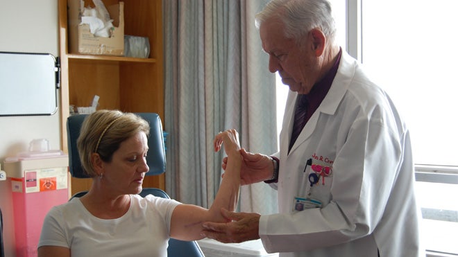
At this point, Madsen feared she would lose her arm – or even worse – her life. Finally, she was introduced to Dr. John Crew, a vascular surgeon and wound specialist at Seton Medical Center in Daily City, Calif., where she was receiving treatment. Crew told Madsen he might know what was causing her health problems: A deadly disease known as necrotizing fasciitis.

The flesh-eating disease

Necrotizing fasciitis, commonly known as the flesh-eating disease, results from a bacterial infection and rapidly destroys the body’s soft tissue. The condition garnered national attention in 2012, when 24-year-old Aimee Copeland underwent a quadruple amputation after contracting necrotizing fasciitis in the aftermath of a zip lining accident.

Typically, necrotizing fasciitis is treated with antibiotics and surgical excision of the infected areas of the body. Though rare, the disease can carry a fatality rate of up to 70 percent – and those that survive are often left with devastating handicaps due to loss of limbs.

“They excise (the dead tissue), and (sometimes) you excise the hands and the legs and that’s a lousy way to end up,” Crew said.

Desperate to save Madsen’s limbs and life, Crew, director of the hospital’s Advanced Wound Care Center, devised a plan in which he would excise the dead tissue from Madsen’s arm and then regularly irrigate the area with an FDA-approved wound cleanser called NeutroPhase. Crew is a paid consultant for NovaBay Pharmaceuticals, the company that manufactures NeutroPhase, and he had been using the product to sterilize wounds for many years. NeutroPhase contains hypochlorous acid, a common chemical disinfectant.

“Hypochlorous acid is produced by the body’s white blood cells when it fights infection,” Dr. Harvey Himel, medical director of the wound program at Icahn School of Medicine at Mount Sinai in New York City, told FoxNews.com. “(It) is one of the common chemicals found to purify water in swimming pools and is used as a disinfectant in food preparation.”  Himel was familiar with the study, but not involved in Madsen’s treatment.

Luckily, Madsen’s initial surgical treatment –coupled with the NeutroPhase irrigation – appeared successful.

However, six days later, Crew noticed another infected spot in a different area on Madsen’s arm. This time, Crew decided to simply insert a catheter and irrigate the area with NeutroPhase – without performing surgery to excise any more of the tissue in her arm.

Remarkably, this area of Madsen’s arm healed just as quickly as the area that underwent the standard surgical excision. Additionally, using NeutroPhase bypassed the severe scarring that now covered much of the rest of her limb.

Madsen noticed a difference in her condition almost immediately after being treated by Crew.

“Before they started NeutroPhase, the pain was unbearable. You can’t describe the way the pain is, and the fever I had was just unbelievable,” Madsen said. “But then, after they started the NeutroPhase and started killing all of the toxins in my arm, the fever subsided and went away. The pain wasn’t as bad…It wasn’t the kind of pain that you feel when it’s infected, and your arm is dying.”

‘People don’t have to lose their limbs or their lives’

Madsen eventually made a full recovery, and while she sustained some nerve damage in her arm, she has regained full function in the limb and now lives a normal life.

After Madsen’s recovery, Crew set out to discover what it was about NeutroPhase that had halted the infection.

“We had to go back to the lab after Lori was healed,” Crew said. “They isolated five or six of the toxins involved in this kind of necrotizing fasciitis, and individually, they treated cells in the lab… and it killed them.”

Crew and his fellow researchers discovered that NeutroPhase seemed to effectively neutralize the toxins produced by the infection, halting the body’s inflammatory-reaction and allowing the patient to begin to heal normally.

Crew recently published his findings in the peer-reviewed journal,Wounds, and he hopes to convince other doctors to begin using NeutroPhase to treat necrotizing fasciitis. Since Madsen’s case, Crew said he has successfully treated several other patients with necrotizing fasciitis using NeutroPhase – even avoiding surgery, in some cases.

“I had one 95-year-old, (and) when I just put in a catheter and irrigated it with NeutroPhase, she healed from that standpoint,” Crew said. “We didn’t need a big massive operation to drain or excise necrotic tissue. We’re looking to tell people this is the way to treat this problem. We won’t make big massive incisions, but small incisions to get the irrigation going as quick as we can.”

Himel warned that while this case appeared to be successful, more research is still needed.

“Since this is a single case report, it is hard to say if this treatment was instrumental in the patient’s recovery,” Himel said. “In order to scientifically prove the value of this additional treatment, they would need to conduct more extensive research.”

For Madsen, her hope is that this treatment will eventually help prevent others in her situation from going through the same agony she did.

“I don’t want to see anyone go through what I went through. I want the word out there that this stuff works on this necrotizing fasciitis,” Madsen said. “People don’t have to lose their limbs or their lives.”

Source: inagist


Babies Seem To Know Themselves Soon After Birth

Understanding you exist as a person happens a lot sooner than you might think.

A study involving 40 cute, pudgy babies found that they were aware of their bodies — and even displayed a sense of ownership of them — less than two days after being born.

Both of those qualities are key ingredients in realizing your own existence, says the study’s lead author, Maria Laura Filippetti, a doctoral candidate specializing in cognitive development at Birkbeck College, University of London.

“Body awareness refers to the feeling of being alive,” she told Shots. “Body ownership refers to the feeling of having a body, the sense that this body belongs to me.”

Past studies reveled how important these two aspects of human life were for infants, but this study was the first to discover it in newborns at birth.

How did the researchers figure it out? Filippetti and her colleagues tested the infants’ ability to recognize themselves using a test similar to the old rubber hand illusion.

That test tricks the mind into thinking a fake rubber hand actually belongs to a person’s body. Researchers lightly stroke a person’s real hand with a paintbrush while it’s hidden from his or her view. Simultaneously, the researchers stroke a rubber hand that’s in plain sight. Stroking the two at the same time and in the same places means the person feels the paintbrush while seeing the action elsewhere.

Normally, a person’s brain associates the feeling of one’s hand with the sight of the hand. But the brain can be confused by a trick like this and start to think the rubber hand is the one it should pay attention to.

For the infants, the test was very similar. Again, a paintbrush was used, but this time the researchers stroked the babies’ cheeks as they watched a video of the same thing happening to another baby.

The researchers tested how the babies behaved when the paintbrush was touched at different times and at the same time on their faces. Since babies can’t talk, their researchers gauged the babies’ reactions by measuring how long they looked at the baby in the video, Filippetti says.

“A longer looking time for a stimulus compared to another one is a measure of discrimination and preference for that stimulus,” she tells Shots.

The newborns did watch the other baby in the video longer when the paintbrush strokes on both happened simultaneously, rather than at different times, or not at all. That response to simultaneous stimulation shows a sense of body awareness and ownership, the researchers say. Here’s a video of how the test went.

The researcher also performed a second experiment with a twist: They showed the babies the same video turned upside down. The babies tested didn’t respond to the simultaneous paintbrush strokes.

The study was published Thursday in Current Biology.

Filippetti concedes that her tests don’t prove with absolute certainty that the infants identified themselves. But she says the work suggests that “the same factors known to be involved in body awareness in adults are present at birth.”

Filippetti says that understanding the typical development of self could someday lead to insights into atypical developments, such as autism spectrum disorder.

For the babies in the experiment, a long nap was probably a richer reward than any contribution to science they might have made.

Source: NPR


Mini-Kidney’ Structures Generated from Human Stem Cells for First Time

Diseases affecting the kidneys represent a major and unsolved health issue worldwide. The kidneys rarely recover function once they are damaged by disease, highlighting the urgent need for better knowledge of kidney development and physiology.

Now, a team of researchers led by scientists at the Salk Institute for Biological Studies has developed a novel platform to study kidney diseases, opening new avenues for the future application of regenerative medicine strategies to help restore kidney function.

For the first time, the Salk researchers have generated three-dimensional kidney structures from human stem cells, opening new avenues for studying the development and diseases of the kidneys and to the discovery of new drugs that target human kidney cells. The findings were reported November 17 in Nature Cell Biology.

Scientists had created precursors of kidney cells using stem cells as recently as this past summer, but the Salk team was the first to coax human stem cells into forming three-dimensional cellular structures similar to those found in our kidneys.

“Attempts to differentiate human stem cells into renal cells have had limited success,” says senior study author Juan Carlos Izpisua Belmonte, a professor in Salk’s Gene Expression Laboratory and holder of the Roger Guillemin Chair. “We have developed a simple and efficient method that allows for the differentiation of human stem cells into well-organized 3D structures of the ureteric bud (UB), which later develops into the collecting duct system.”

The Salk findings demonstrate for the first time that pluripotent stem cells (PSCs) — cells capable of differentiating into the many cells and tissue types that make up the body — can made to develop into cells similar to those found in the ureteric bud, an early developmental structure of the kidneys, and then be further differentiated into three-dimensional structures in organ cultures. UB cells form the early stages of the human urinary and reproductive organs during development and later develop into a conduit for urine drainage from the kidneys. The scientists accomplished this with both human embryonic stem cells and induced pluripotent stem cells (iPSCs), human cells from the skin that have been reprogrammed into their pluripotent state.

After generating iPSCs that demonstrated pluripotent properties and were able to differentiate into mesoderm, a germ cell layer from which the kidneys develop, the researchers made use of growth factors known to be essential during the natural development of our kidneys for the culturing of both iPSCs and embryonic stem cells. The combination of signals from these growth factors, molecules that guide the differentiation of stem cells into specific tissues, was sufficient to commit the cells toward progenitors that exhibit clear characteristics of renal cells in only four days.

The researchers then guided these cells to further differentiated into organ structures similar to those found in the ureteric bud by culturing them with kidney cells from mice. This demonstrated that the mouse cells were able to provide the appropriate developmental cues to allow human stem cells to form three-dimensional structures of the kidney.

In addition, Izpisua Belmonte’s team tested their protocol on iPSCs from a patient clinically diagnosed with polycystic kidney disease (PKD), a genetic disorder characterized by multiple, fluid-filled cysts that can lead to decreased kidney function and kidney failure. They found that their methodology could produce kidney structures from patient-derived iPSCs.

Because of the many clinical manifestations of the disease, neither gene- nor antibody-based therapies are realistic approaches for treating PKD. The Salk team’s technique might help circumvent this obstacle and provide a reliable platform for pharmaceutical companies and other investigators studying drug-based therapeutics for PKD and other kidney diseases.

“Our differentiation strategies represent the cornerstone of disease modeling and drug discovery studies,” says lead study author Ignacio Sancho-Martinez, a research associate in Izpisua Belmonte’s laboratory. “Our observations will help guide future studies on the precise cellular implications that PKD might play in the context of kidney development.”

Source: Science Daily


Genetically engineered tomatoes could help improve cholesterol levels

Researchers have reported that small amounts of a specific type oflipid in the small intestine could play a greater role than earlier thought in generating the high cholesterol levels and inflammation that lead to cloggedarteries.

The tomatoes, created at UCLA, produce a small peptide called 6F that mimics the action of apoA-1, the chief protein inHDL.

Researchers added 2.2 percent (by weight) of freeze-dried tomato powder from the peptide-enhanced tomatoes to low-fat, low-cholesterol mouse chow that was supplemented with LPAs.

They also added the same dose of the peptide-enhanced tomatoes to the high-fat high- cholesterol diet.

They found that this addition to both diets prevented an increase in the level of LPAs in the small intestine and also stopped increases in “bad” cholesterol, decreases in “good” cholesterol and systemic inflammation. Tomatoes that did not contain the peptide had no effect.

According to senior author Dr. Alan Fogelman, executive chair of the department of medicine and director of the atherosclerosis research unit at the David Geffen School of Medicine at UCLA, the peptide-enhanced tomatoes may work in large part by reducing the amount of the LPAs in the small intestine.

The study has been published in the Journal of Lipid Research.

Source: News track India


Car mechanic invents new device to aid in childbirth

Health experts say the Odon Device has the potential to save lives around the world.

Argentinean Jorge Odon is a car mechanic by trade, and a tinkerer by nature. Recently, Odon watched a video about an easy method for removing a cork stuck in a wine bottle. And in the middle of the night it dawned on him that the same “trick” could be used during childbirth to help a baby that is stuck in the birth canal.

Obstructed labor — when the baby’s head gets stuck in the birth canal — is a major complication of childbirth. Doctors may use forceps or suction cups to try to pull the baby out. These procedures can lead to a number of complications on their own, and still are not guaranteed to succeed. In wealthier countries, the mother and baby may be whisked off to the operating room for an emergency C-section. In poor countries, or communities without access to advanced health care, this type of surgery is not an option.

Odon’s children were fortunately born without complications, but his aunt suffered nerve damage during childbirth, so Odon was familiar with the potential complications. In an interview with the New York Times, Odon explained that after seeing the wine bottle trick, it dawned on him that this could be used during childbirth.

With the help of his wife, he constructed a prototype using his daughter’s baby doll, a glass jar and a fabric bag.

In time, and with several revisions of his design, Odon’s idea — the Odon Device — won the endorsement of the World Health Organization (WHO), big-time donors, and a medical technology company that wants to develop it for production.

Here’s how it works:

Using the Odon Device, a lubricated plastic sleeve is slipped around the baby’s head and inflated until it forms a grip. Doctors then pull on the bag until the baby emerges. According to Dr. Margaret Chan, director general of WHO, the Odon Device has the potential to save babies in poor countries, and reduce the number of emergency cesareans in rich ones.

“The Odon Device, developed by WHO and now undergoing clinical trials, offers a low-cost simplified way to deliver babies, and protect mothers, when labour is prolonged. It promises to transfer life-saving capacity to rural health posts, which almost never have the facilities and staff to perform a C-section. If approved, the Odon Device will be the first simple new tool for assisted delivery since forceps and vacuum extractors were introduced centuries ago,” Chan said in a speech to the 65th World Health Assembly.

Source: mnn.com


Philanthropist’s gift a big bang for stem cell research

A philanthropist who made his money as a credit card provider is giving $100 million to human stem-cell research.

The money will go to the University of California at San Diego during the next five years as researchers reach certain milestones, said T. Denny Sanford, who founded First Premier Bank here and offers low-limit Master cards and Visas to customers with poor credit through Premier Bankcard. United National Corp., where Sanford is now chief executive, owns both companies.

“This, in my opinion, is the medicine of the future,” he said. “The potential of stem cells is just unbelievable.”

The money will support the hiring of 20 or more scientists and efforts to recruit patients for drug trials along with new construction at the San Diego complex.

The donation pushes Sanford past the $1 billion mark for total gifts to health care and research, he said.

Sanford, 77, has homes in South Dakota, Arizona and California. On Oct. 19, he suffered a pulmonary embolism — a blood clot in the lungs — while on a hunting trip with friends near Gregory, S.D., about 140 miles west of here.

He said he was saved because of a middle-of-the-night medical flight to Sanford University of South Dakota Medical Center here, helicopter and plane flights made possible in part because of donations he has made to what is now Sanford Health system.

“I was within minutes or hours of death,” he said. His physician here, Dr. Eric Larson, said Sanford is doing fantastic, playing golf regularly and exercising on an elliptical machine, less than a month after getting clot-busting medications to treat the condition.

Most of Sanford’s donations, about $700 million, have gone to the Sanford Health. He has pledged to give all his money away. He said he still has close to $1 billion.

The $100 million he is committing to UC San Diego is the lead resource in a project that officials say will cost a total of $275 million.

What now is called the Sanford Consortium for Regenerative Medicine includes scientists from five institutions — UC San Diego, Sanford-Burnham, Scripps, the Salk Institute for Biological Studies and the LaJolla Institute for Allergy and Immunology.

“Overall, the effort is to bring stem cell research into human clinical trials,” said Debra Kain, director of health sciences research communications at UC San Diego.

In Sioux Falls, Sanford made a $400 million donation to the nonprofit medical center in 2007 and established four priorities, one of which was curing a major disease that officials later pegged as Type 1 diabetes. Another of his donations here, $100 million in 2011, is for research and treatment for breast cancer. His mother, Edith, died of the disease when he was 4 years old.

This gift is different because he has no personal or family connection to the neurological diseases he hopes that stem cell research can address.

Research so far has been instructive on the use of mice and monkeys, so it’s time now to extend the effort to humans, he said.

“We are excited about some major potential cures, particularly with neurological diseases like Lou Gehrig’s disease, or spinal cord injuries,” Sanford said. Lou Gehrig’s disease, also called amyotrophic lateral sclerosis, affects nerve cells in the brain and spinal cord that control muscle movement. It has no cure.

Sanford is excited that the work could lead several directions.

“It could be spinal cord injuries, Alzheimer’s, Parkinson’s, eventually heart and cancer and diabetes,” he said.

Source: USA Today


IBM opens global research lab in Africa IBM’s

12th global research lab was opened here last week. It is designed to conduct applied and far-reaching exploratory research into the big challenges of the African continent and deliver commercially viable innovations that impact people’s lives.

 The facility features one of Africa’s most powerful computer hubs, giving IBM researchers the ability to analyse and draw insight from vast amounts of data in search for solutions to Africa’s challenges such as energy, water, transportation, agriculture, healthcare, financial inclusion, human mobility and public safety.

“The establishment of this research laboratory underpins the government’s commitment to innovation ecosystems that are already available in Kenya,” said the president of Kenya, Uhuru Kenyatta. The lab is supported by the Kenyan ICT Authority.

The lab’s research agenda will include the development of cognitive computing technologies that integrate learning and reasoning capabilities, enabling experts to make better decisions in areas such as healthcare delivery and financial services.

“We are currently experiencing the emergence of a new Africa – one where science and technology are enabling a pivotal ‘leap frog’ moment allowing governments and businesses to drive economic growth, raise the standard of living and compete with their global counterparts,” said Kamal Bhattacharya, director, IBM Research-Africa.

“The launch of Africa’s first full-scale, technology research facility will help lay the foundation for the continent’s future scientific and economic independence,” he added. Nicholas Nesbitt, country general manager, East Africa, said it was not just about science and technology, “but also about innovating new business models and partnering with local enterprises to ensure that our new solutions have the maximum impact on business and society.”

Source: The Times of India


New imaging method ‘predicts’ heart attack risk

The Centers for Disease Control and Prevention (CDC) states that around 715,000 Americans suffer a heart attack every year. Now, scientists have created a new imaging technique that could identify which patients are at high risk. This is according to a study published in the The Lancet.
Researchers from the University of Edinburgh in the UK say the test – carried out using positron emission tomography (PET) and computed tomography (CT) – is able to “light up” dangerous fatty plaques in the arteries that are in danger of rupturing. This is a process that can cause heart attacks.

To reach their findings, the researchers analyzed 80 patients. Of these, 40 patients recently had a heart attack, while the other 40 patients had angina – restricted blood supply to the heart posing a higher risk of heart attack.

‘First step’ towards heart attack prevention

Using the PET-CT scanner, the researchers found that 90% of patients who had a heart attack showed a “lit up” yellow area in one of their blood vessels. This area corresponded exactly to the location of the plaque that caused the patients’ heart attacks, the researchers say.

The scanner also showed lit up plaques in around 40% of the patients with angina. Furthermore, the researchers found “high-risk” features in these patients that suggested a heart attack may be imminent, meaning they were in need of aggressive drug treatment or surgery.

Dr. Marc Dweck, of the University of Edinburgh and lead study author, says their findings are a step toward heart attack prevention:

“We have developed what we hope is a way to ‘light up’ plaques on the brink of rupturing and causing a heart attack.

If we could know how close a person is to having a heart attack, we could step in with medication or surgery before the damage is done. This is a first step towards that goal.”

Potential for identifying ‘ticking time bomb’ patients

The researchers say the next stage of this research is to confirm the findings and to determine whether the PET-CT imaging technique can improve the management and treatment of patients with coronary artery disease.

Prof. Peter Weissburg, medical director at the British Heart Foundation in the UK, which part-funded the study, notes that the technique looks promising:

“Being able to identify dangerous fatty plaques likely to cause a heart attack is something that conventional heart tests can’t do. This research suggests that PET-CT scanning may provide an answer – identifying ‘ticking time bomb’ patients at risk of a heart attack.”

“We now need to confirm these findings, and then understand how best to use new tests like this in the clinic to benefit heart patients,” he adds.

Source: Medical News Today


How brain uses sleep for visual task learning

Particular frequencies to consolidate learning in specific brain regions.

 In August, Brown University scientists reported that two specific frequencies, fast-sigma and delta, that operated in the supplementary motor area of the brain were directly associated with learning a finger-tapping task akin to typing or playing the piano.

The new results show something similar with a visual task in which 15 volunteers were trained to spot a hidden texture amid an obscuring pattern of lines.Takeo Watanabe, professor of cognitive, linguistic, and psychological sciences at Brown, said that perceptual learning in general has been found to improve the visual ability of patients who have some decline of function due to aging.

In this case the researchers, led by graduate student Ji Won Bang, devised an experiment to see how sleep may help such training take hold. They measured the brainwaves of the participants during sleep before and the training, and they measured the volunteers’ performance on the task before and after.

The researchers saw significant increases in sigma brainwave power after sleep compared to before in the visual cortical area in the occipital lobe of the volunteers’ brains.

To ensure they were measuring activity related to learning the task, the researchers purposely put the stimulus of the discrimination task in a particular quadrant of the subjects’ field of view.

That position corresponds to an anatomically distinct part of the visual cortical area. The team saw that the measured gain in sigma wave power was greater specifically in that trained part of the visual cortical area rather than in the untrained parts.

They also saw that the difference of power increase between trained and untrained regions of the visual cortical area was correlated with each individual’s performance improvement on the task.

The study has been published in the Journal of Neuroscience.

Source: News Track India