Cause of infant deaths from SIDS identified

Researchers have claimed that babies dying from Sudden infant death syndrome (SIDS) have brain stem abnormalities regardless of whether they were exposed to risks like suffocation or co-sleeping.

The researchers analysed the brain stems of 71 infants who had died suddenly and unexpectedly over 11 years.
The study found that all the babies who died had abnormalities of four neurochemicals in the brain stem, located at the skull’s base and connects the brain to the spinal cord.

According to Boston Children’s Hospital and Florey Institute of Neuroscience and Mental Health’s Dr Jhodie Duncan, the research suggests that the abnormality leaves the toddlers unable to adequately respond when faced with a stressor while sleeping.
He said that if a pillow goes over a healthy infant’s face, their brain usually detects changes in oxygen levels and initiates response, so that the baby can turn its head and continue breathing. However, babies with the abnormality did not “respond properly” in the same situation, which lead to their death, News.com.au reported.

The next possible step of the research team would be to see if a blood test can be developed to be used as an early screening tool to identify infants at risk of sudden and unexpected death in their first year.
The research has been published in the journal Pediatrics.

Source: Yahoo news


Study shows how brain forms memories

Scientists have discovered how memories are stored in specific brain cells. The new study also pinpoints how these incidents are recalled.

Using a video game in which people navigate through a virtual town delivering objects to specific locations, a team of neuroscientists from the University of Pennsylvania and Freiburg University has discovered how brain cells that encode spatial information form “geotags” for specific memories, and are activated immediately before those memories are recalled.

Their work showed how spatial information is incorporated into memories, and why remembering an experience can quickly bring other events to mind that happened in the same place, reports the Science Daily.

“These findings provide the first direct neural evidence for the idea that the human memory system tags memories with information about where and when they were formed, and that the act of recall involves the reinstatement of these tags,” said Michael Kahana, professor of psychology in Penn’s School of Arts and Sciences.

Source: Daijiworld


Research: first functional lung and airway cells from stem cells

Scientists have succeeded in transforming human stem cells into functional lung and airway cells, thus giving way to the possibility of generating lung tissue for transplant using a patient’s own cells.

The study by Columbia University Medical Center (CUMC) researchers has significant potential for modelling lung disease, screening drugs, studying human lung development, and, ultimately, generating lung tissue for transplantation.

“Researchers have had relative success in turning human stem cells into heart cells, pancreatic beta cells, intestinal cells, liver cells, and nerve cells, raising all sorts of possibilities for regenerative medicine,” study leader Hans-Willem Snoeck said.

“Now, we are finally able to make lung and airway cells. This is important because lung transplants have a particularly poor prognosis. Although any clinical application is still many years away, we can begin thinking about making autologous lung transplants-that is, transplants that use a patient’s own skin cells to generate functional lung tissue.”

The research builds on Dr. Snoeck’s 2011 discovery of a set of chemical factors that can turn human embryonic stem (ES) cells or human induced pluripotent stem (iPS) cells into anterior foregut endoderm-precursors of lung and airway cells.

The findings have implications for the study of a number of lung diseases, including idiopathic pulmonary fibrosis (IPF), in which type 2 alveolar epithelial cells are thought to play a central role.

The study was published in the journal Nature Biotechnology.

Source: News Track India


New treatment for osteoporosis discovered

After more than four years of investigation, researchers from the Ageing Bone Research Program (Sydney Medical School’s Nepean campus), have found the treatment has shown very promising results in animal experiments.

The compound is called picolinic acid, a product derived of the essential amino acid tryptophan.

Lead researcher Professor Gustavo Duque said the odorless compound can be easily dissolved in water.

“This is a major step in the development of a completely new type of medication for osteoporosis. Instead of stopping bone destruction, our compound instead stimulates bone formation,” he said.

“The product is easily dissolved in water, has a higher level of absorption and did not induce any side effects in the treated mice.

“When this medication was administered in the water of normal and menopausal mice, picolinic acid strongly and safely increased bone mass in normal mice and rescued bone from menopause-associated osteoporosis.”

Professor Duque said the team had patented the compound and will expand their trials to humans in the near future in a bid to address the increasing numbers of people developing the condition.

“Osteoporosis affects an estimated 300 million people worldwide. One in three women over 50 will experience osteoporotic fractures, as will one in five men.

“Despite the current treatments available, by 2050, the worldwide incidence of hip fracture in men is projected to increase by 310 percent and 240 percent in women.

“This increase is explained by the low rate of diagnosis and treatment for osteoporosis, as well as some concerns about the potential side effects of the current treatments.

“There are also close similarities between the majority of the osteoporosis medications in terms of their anti-fracture effect and mechanism of action.”

According to Professor Duque, there is a reduction in bone formation as part of the ageing process that predisposes people to osteoporosis.

“In this case we are targeting the real problem by stimulating the bone forming cells to work and produce more bone, thus increasing bone mass and hopefully preventing new fractures,” he said.

Source: The University of Sydney


GE unveils ultra high frequency mobile X-ray

GE mobile xrayBengaluru: GE Healthcare on Thursday announced the launch of Brivo XR115, a next-generation ultra high frequency (200KHz) mobile X-ray system designed and developed in India for the world. The new GE Brivo XR 115 addresses five critical problems faced by radiologists and radiographers — image quality from mobile machines of critical anatomies like the spine, patient discomfort which can occur while shifting from hospital bed to the X ray room, higher radiation uncontrolled patient movement that occurs during paediatric imaging, and the potential safety issues that can exist moving equipment between wards, according to a statement by GE Healthcare.

X-ray is the primary and most widely used imaging tool in healthcare. India is home to about 100,000 traditional X-ray systems producing an estimated 100 billion X-ray images a year. The majority of these X-ray systems are old generation technology plagued by critical problems.

GE’s new Brivo XR115 ultra high frequency 100 ma mobile X ray provides a solution with great images of all anatomies, lower radiation dose on account of ultra high frequency technology, freedom of mobility for its light weight design, lower maintenance cost due to safe design and, the added peace of mind for radiographers, radiologists and patients, the statement said.

“We are attempting to address the severe challenges faced by Indian healthcare providers by building a healthier India through technology innovation. Our customers asked us to make X-ray imaging, the first step in better diagnosis safer, more affordable and accessible. Here we are, with Brivo XR 115 addressing all their challenges and helping build a healthier India,” said Terri Bresenham, president and CEO, GE Healthcare, South Asia.

“The Brivo XR115 will bring complete peace of mind to the radiologist, radiographer and patient alike. Keeping in mind the concerns of radiologists and physicians, our research team and designers have created a safer imaging environment by reducing X-ray exposure time, lowering radiation dose and providing exceptional images,” said Shireesh Sahai, director, DGS (detection and guidance solutions) and surgery, GE Healthcare, South Asia.

Source: India Medical Times


New drug target to help fight malaria identified

A team of researchers has identified a key metabolic enzyme that is required by the common malaria parasites at all stages of its life cycle for survival in humans.

Co-first author Marcus C. S. Lee, PhD, associate research scientist in microbiology and immunology at Columbia University Medical Center (CUMC) said the study is important because most anti malarials are effective at killing the parasites only as they circulate in the bloodstream. However, the parasites can hide in the liver for years before reemerging and triggering a relapse of the disease.

The other co-first author is Case W. McNamara, PhD, research investigator at the Genomics Institute for the Novartis Research Foundation. The study leaders are Elizabeth A. Winzeler, PhD, professor of pharmacology and drug discovery at University of California San Diego, and Thierry Diagana, head of Novartis Institute for Tropical Diseases in Singapore.

The enzyme, phosphatidylinositol 4-kinase (PI4K), was found by screening more than a million drug compounds against Plasmodium falciparum, the parasite responsible for the most lethal form of malaria. Using this screen, the researchers found a class of compounds known as imidazopyrazines, which are capable of killing several species of Plasmodium at each stage of the parasites’ life cycle in its vertebrate host.

The researchers identified the target of the imidazopyrazines by evolving parasite cell lines that were resistant against the drugs and then analyzing the parasites’ genomes for the changes responsible for conferring resistance. Those genetic changes pointed to the gene that encodes PI4K.

The CUMC team, led by David Fidock, PhD, professor of microbiology and immunology and medical sciences (in medicine), used novel genetic tools to confirm that PI4K was being directly targeted by the imidazopyrazines.

Then, using cellular imaging, it was found that imidazopyrazines interfere with the function of PI4K on the parasite Golgi (the organelle that packages proteins for delivery to other cellular destinations). ”

The study is published in the journal Nature.

Source: top news


Honey bees can be trained to detect cancer

Dezeen_Susana_Soares_Bees_Design_2

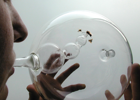
Portuguese designer Susana Soares has developed a device for detecting cancer and other serious diseases using trained bees

The bees are placed in a glass chamber into which the patient exhales; the bees fly into a smaller secondary chamber if they detect cancer.

“Trained bees only rush into the smaller chamber if they can detect the odour on the patient’s breath that they have been trained to target,” explained Soares, who presented her Bee’s project at Dutch Design Week in Eindhoven last month.

Scientists have found that honey bees – Apis mellifera – have an extraordinary sense of smell that is more acute than that of a sniffer dog and can detect airborne molecules in the parts-per-trillion range.

Bees can be trained to detect specific chemical odours, including the biomarkers associated with diseases such as tuberculosis, lung, skin and pancreatic cancer.

Bees have also been trained to detect explosives and a company called Insectinl is training “sniffer bees” to work in counter-terrorist operations.

“The bees can be trained within 10 minutes,” explains Soares. “Training simply consists of exposing the bees to a specific odour and then feeding them with a solution of water and sugar, therefore they associate that odour with a food reward.”

Once trained, the bees will remember the odour for their entire lives, provided they are always rewarded with sugar. Bees live for six weeks on average.

“There’s plenty of interest in the project especially from charities and further applications as a cost effective early detection of illness, specifically in developing countries,” Soares said.

Bee’s explores how we might co-habit with natural biological systems and use their potential to increase our perceptive abilities.Dezeen_Susana_Soares_Bees_Design_3
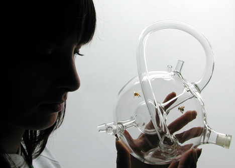
The objects facilitate bees’ odour detection abilities in human breath. Bees can be trained within 10 minutes using Pavlov’s reflex to target a wide range of natural and man-made chemicals and odours, including the biomarkers associated with certain diseases.

The aim of the project is to develop upon current technological research by using design to translate the outcome into systems and objects that people can understand and use, engendering significant adjustments in their lives and mind set.

How it works

The glass objects have two enclosures: a smaller chamber that serves as the diagnosis space and a bigger chamber where previously trained bees are kept for the short period of time necessary for them to detect general health. People exhale into the smaller chamber and the bees rush into it if they detect on the breath the odour that they where trained to target.

What can bees detect?

Scientific research demonstrated that bees can diagnose accurately at an early stage a vast variety of diseases, such as: tuberculosis, lung and skin cancer, and diabetes.

Precise object

The outer curved tube helps bees avoid from flying accidentally into the interior diagnosis chamber, making for a more precise result. The tubes connected to the small chamber create condensation, so that exhalation is visible.

Detecting chemicals in the axilla

Apocrine glands are known to contain pheromones that retain information about a person’s health that bee’s antennae can identify.

The bee clinic

These diagnostic tools would be part of system that uses bees as a biosensor.

The systems implies:
– A bee centre: a structure that facilitates the technologic potential of bees. Within the centre is a bee farm, a training centre, a research lab and a health care centre.

– Training centre: courses can be taken on bee training where bees are collected and trained by bee trainers. These are specialists that learn bee training techniques to be used in a large scope of applications, including diagnosing diseases.

– BEE clinic: bees are used at the clinic for screening tests. These insects are very accurate in early medical diagnosis through detection on a person’s breath. Bees are a sustainable and valuable resource. After performing the  diagnose in the clinic they are released, returning to their beehive.

Bee training

Bees can be easily trained using Pavlov’s reflex to target a wide range of natural and man-made chemicals odours including the biomarkers associated with certain diseases. The training consists in baffling the bees with a specific odour and feeding them with a solution of water and sugar, therefore they associate that odour with a food reward

Source: de zeen magazine


New technique to treat parasitic cystic tumour of kidney

Dr Santosh Kumar, assistant professor, department of urology, Postgraduate Institute of Medical Education and Research (PGIMER), Chandigarh, has developed an innovative surgical technique to treat parasitic cystic tumour of kidney, a rare disease that can lead to destruction of kidney.

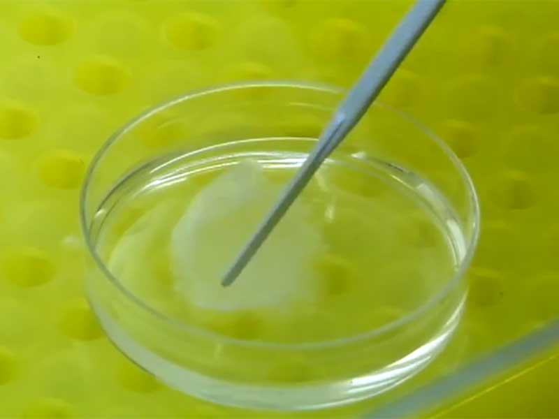
In this Santosh PGI Technique, Dr Kumar operated a 22-year-old woman by single 2 cm incision through umbilicus, the natural scar given by god, using conventional port and instrument.

The innovative surgical technique is described in a paper published in the Asian Journal of Endoscopic Surgery, a journal of the Japan Society of Endoscopic Surgeons.

According to Dr Kumar, the new minimal invasive technique helped the patient recover fast and without scar. Her kidney was saved. “We believe it is the first case of this kind in this large hydatid parasitic tumour in which this technique was used,” he said.

Parasitic cystic tumour is common in canine, dogs, sheep etc. Humans get infected by eggs and embryo in contaminated vegetable and meat. The patient may not have any symptoms, can present with palpable tumour in abdomen, allergic reaction and obstruction of kidney, according to a statement by PGIMER.

Rupture of parasitic cystic tumour like mass can lead to shock and death. Removal of kidney by open surgery used to be traditional treatment, the statement said.

Treating a giant parasitic tumour kidney was earlier reported by Dr Kumar in the Journal of Endourology, an American Endourological Society journal. In this article he described how he performed treatment of various genitourinary hydatid cystic mass by laparoscopy by three small incisions.

Talking about his latest innovation, Dr Kumar said, “In our case the patient was newly married, who had been deserted by her husband because of unfortunate social condition and risk of morbidity of disease and surgery. Single hole surgery with this new technique through natural scar i.e. umbilicus guided by nephroscope, which is very common armamentarium of urologist, was an excellent advantageous condition for her.”

“It is interesting to say that after successful surgery the husband united with her wife happily,” he added.

Source: India Medical Times


Thinking too much about food could make us eat more

A new analysis of 50 studies has found that thinking before eating may actually undermine people’s dieting goals.

Jessie De Witt Huberts of Utrecht University says that we are expert rationalizers when it comes to finding a reason to eat more. He said that people seem to

be very creative in coming up with such reasons, asserting that they can justify having the cake on account that it has been a hard day, or that they will exercise tomorrow, that it is a special occasion, or that it is impolite to refuse.

Huberts said that this is when justification processes become a slippery slope – as the reasons are often applied ad hoc, they no longer form strict rules that regulate when you stick to your diet and when you can cut yourself some slack.

A growing body of research has found that such justification can even come from performing well on a task or from doing good for others. In several studies, people who received positive feedback on a task were more likely to choose an unhealthy versus a healthy option.

Source: Hindustan Times


Artificial skin created using stem cells from umbilical cord

In a first, scientists have grown artificial skin using stem cells derived from the umbilical cord.

Scientists from the Tissue Engineering Research Group at the Department of Histology at the University of Granada demonstrated the ability of Wharton jelly mesenschymal stem cells to turn to oral-mucosa or skin-regeneration epithelia.

To grow the artificial skin, the researchers used, in addition to this new type of epithelia covering, a biomaterial made of fibrin and agarose, already designed and developed by the University of Granada research team.

Prior studies from the same team, already pointed to the possibility that Wharton stem cells could be turned into epithelia cells.

The current work is the confirmation of those initial studies and its application to two regeneration structures: skin and oral mucosa, increasingly needed in injuries in these parts of the body.

One of the problems major-burn victims currently have is that, in order to apply the current techniques of artificial skin, a number of weeks are needed. That is because the skin needs to be grown from parts of the patient’s healthy skin.

“Creating this new type of skin using stem cells, which can be stored in tissue banks, means that it can be used instantly when injuries are caused, and which would bring the application of artificial skin forward many weeks,” said Antonio Campos, Professor of Histology at the University of Granada and one of the authors of this study.

The study is published in the journal Stem Cells Translational Medicine.

Source:  The Hindu