New lung cancer screening guidelines approved for older smokers

Guidelines recommending annual low-dose CT lung cancer screening for older smokers have been approved by the US Preventive Services Task Force. The recommendations apply to individuals aged between 55 and 80 who are at high risk for lung cancer as a result of heavy smoking.

The guidelines are published in the journal Annals of Internal Medicine.

According to the American Cancer Society, approximately 228,190 new cases of lung cancer will have been diagnosed during 2013, with 159,480 deaths from the disease. This accounts for around 27% of all cancer deaths.

Background information from the guidelines states that around 85% of all cases of lung cancer are caused by smoking, and the risk of lung cancer increases with age, particularly for those aged over 55.

Dr. Michael LeFevre, co-vice chair of the US Preventive Services Task Force (USPSTF), says these factors suggest that the longer a person smokes, the higher their risk is for developing lung cancer.

He adds:

“When clinicians are determining who would most benefit from screening, they need to look at a person’s age, overall health, how much the person has smoked, and whether the person is still smoking or how many years it has been since the person quit.”

Low-dose CT scanning ‘more accurate’
The 2004 lung cancer screening recommendation from the USPSTF stated that the “evidence was insufficient to recommend for or against screening for lung cancer in asymptomatic persons with LDCT (low-dose computed tomography), chest radiography, sputum cytologic evaluation or a combination of these tests.”

With the aim of updating these recommendations, a panel from the USPSTF reviewed more than 33 studies involving current or former smokers who were at average or high risk for developing lung cancer.

The analysis included a study of more than 50,000 people who were a part of the National Lung Screening Trial.

From their research, the panel found that low-dose computed tomography (CT) lung cancer screening was more accurate in identifying the disease in its early stages, compared with alternative screening tests.

Their findings have led the USPSTF to “recommend annual screening for lung cancer with low-dose computed tomography in adults aged 55 to 80 years who have a 30 pack-year smoking history and currently smoke or have quit within the past 15 years.”

A 30-pack year is the equivalent to one pack a day for 30 years, or two packs a day for 15 years.

Screening not recommended when smoking ceased for 15 years
However, they note that screening should be stopped once a person has not smoked for 15 years or develops a health problem that shortens life expectancy or the willingness or ability to undergo potential lung surgery.

Dr. Virginia Moyer, chair of USPSTF emphasizes that it is important to assess a patient’s overall health to determine whether screening is appropriate.

“The benefit of screening may be significantly less in people with serious medical problems and there is no benefit in screening someone for whom treatment is not an option,” she says.

“In these people, screening may lead to unintended harms such as unnecessary tests and invasive procedures.”

She also adds that although screening for lung cancer is beneficial, it should not be seen as an alternative to giving up smoking.

Source: medical news today


New breakthrough brings Muscular Dystrophy Defect drug closer to reality

Researchers have revealed an atomic-level view of a genetic defect that causes a form of muscular dystrophy, myotonic dystrophy type 2.
The scientists from The Scripps Research Institute have used this information to design drug candidates with potential to counter those defects and reverse the disease.

“This the first time the structure of the RNA defect that causes this disease has been determined,” lead author Matthew Disney said. “Based on these results, we designed compounds that, even in small amounts, significantly improve disease-associated defects in treated cells.”

Myotonic dystrophy type 2 is a relatively rare form of muscular dystrophy that is somewhat milder than myotonic dystrophy type 1, the most common adult-onset form of the disease.

Both types of myotonic dystrophy are inherited disorders that involve progressive muscle wasting and weakness, and both are caused by a type of genetic defect known as a “RNA repeat expansion,” a series of nucleotides repeated more times than normal in an individual’s genetic code.

The repeat binds to the protein MBNL1, rendering it inactive and resulting in RNA splicing abnormalities-which lead to the disease.
Using information about the RNA’s structure and movement, the scientists were able to design molecules to improve RNA function.

The new findings were confirmed using sophisticated computational models that show precisely how the small molecules interact with and alter the RNA structure over time.
Those predictive models matched what the scientists found in the study-that these new compounds bind to the repeat structure in a predictable and easily reproducible way, attacking the cause of the disease.

The study is published in journal ACS Chemical Biology. (ANI)

Source: Yahoo news


Scientists discover new way of overcoming human stem cell rejection

Human embryonic stem cells have the capacity to differentiate into a variety of cell types, making them a valuable source of transplantable tissue for the treatment of numerous diseases, such as Parkinson’s disease and diabetes.

But there’s one major issue: Embryonic stem cells are often rejected by the human immune system.

Now, researchers from the University of California San Diego may have found an effective way to prevent this rejection in humans. Utilizing a novel humanized mouse model, the scientists have revealed a unique combination of immune suppressing molecules that stop the immune system from attacking the injected stem cells – without shutting the system down completely.

This discovery could ultimately help resolve some of the major problems currently limiting the use of embryonic stem cells for certain conditions, paving the way for the development of more effective human stem cell therapies.

“This is a generic way of immune suppression, so it could potentially be applied not just for stem cells therapies, but for organ transplants as well,” Yang Xu, a professor of biology at UC San Diego and lead author of the study, told FoxNews.com. “It can be very broad.”

Embryonic stem cells are different from the other cells in a patient’s body, making them “allogenic.” This means the immune system will recognize them as foreign agents and attack them.

One way of overcoming this rejection problem is to give patients immunosuppressant drugs, which suppress the entire immune system. While short term use of immunosuppressants has been successful for many organ transplants, embryonic stem cell therapies for chronic diseases require long term use of these drugs – which can often be very toxic and increase the risk of cancer.

“In order for the patient to really use this therapy, they have to decide: Do they want a lifelong use of immunosuppressant drugs, or are they willing to live with the symptoms of their disease,” Xu said.

Source: news.nom


Cholesterol linked to Alzheimer’s protein, unclear why

Patterns of “good” and “bad” cholesterol usually associated with heart risks also predicted the levels of Alzheimer’s-related beta amyloid protein seen in the brains of study participants.

“One of the important themes emerging from dementia research over the past 15 years is that there are intriguing connections between vascular disease and Alzheimer’s disease,” Bruce Reed, who led the research, told Reuters Health by email.

Reed is a professor and associate director of the University of California Davis Alzheimer’s Disease Research Center.

“It has become increasingly clear that what have been traditionally thought of as vascular risk factors – things like hypertension, diabetes and elevated cholesterol – are also risk factors for Alzheimer’s disease,” Reed said.

In previous work, Reed and his colleagues found a connection between overall vascular risk and levels of brain amyloid.

“Amyloid deposition is important because it is widely believed by scientists to be a key event that initiates a chain of events that eventually, years later, results in the dementia of Alzheimer’s disease,” Reed said.

“There was also previous work in cell cultures and with animals that suggested that cholesterol plays an important role in promoting the deposition of amyloid in the brain,” Reed said.

For the new study, published in JAMA Neurology, the researchers examined the cholesterol levels of 74 elderly people who had normal to mildly impaired cognitive function. Researchers also measured brain deposits of beta amyloid protein with positron emission tomography (PET scanning).

They found that on average, participants who had higher levels of the “bad” LDL cholesterol and lower levels of “good” HDL cholesterol also had higher levels of amyloid in the brain.

Elevated LDL-cholesterol is associated with cardiovascular disease while high levels of HDL-cholesterol are thought to protect against heart disease.

“We think this is a very important finding, but as with all novel findings it needs to be replicated,” Reed said. “Assuming that the basic pattern is found in other groups of patients, it is urgent that we try to understand the mechanism(s) behind this finding.”

“Cholesterol in blood and cholesterol in brain are separate ‘pools,’ walled off from one another by the blood brain barrier. We measured cholesterol in blood. So that is one question that needs to be answered – how do cholesterol levels in blood and in brain influence each other,” Reed said.

“And in the brain, it is not entirely understood how changing cholesterol levels might reduce amyloid deposition. We are very interested in the idea that higher HDL (‘good’) cholesterol levels may help the brain more efficiently clear the toxic amyloid at an early stage,” Reed said.

“If those questions were understood we could begin to think about how to change cholesterol levels so as to prevent the buildup of amyloid,” Reed added.

In other studies, some researchers have found evidence that LDL cholesterol-lowering drugs, such as statins, might offer some protection against Alzheimer’s disease, but the results have been inconsistent.

Reed’s team didn’t see any associations between current use of cholesterol medication by the participants and their amyloid levels.

In their report, Reed and his coauthors caution that the study does not prove cholesterol is directly affecting amyloid deposition. For instance, they write, unhealthy cholesterol could be linked to vascular damage, such as small strokes, and those micro-injuries could be the reason for the protein deposits.

He encourages people to follow the advice of their doctor and try to achieve the cholesterol guidelines set by the American Heart Association.

“A remarkable number of people who are alive now will live into their 80s or beyond – the period of highest risk for Alzheimer’s. This study is one more piece of evidence that what we do now can shape our health positively in those years,” Reed said.

Source: ABS CBN news


Scientists use drug to repair a rare birth defect

University of British Columbia and Vancouver Coastal Health scientists have developed a potential cure for a rare eye disease, showing for the first time that a drug can repair a birth defect.

They formulated the drug Ataluren into eye drops, and found that it consistently restored normal vision in mice who had aniridia, a condition that severely limits the vision of about 5,000 people in North America. A small clinical trial with children and teens is expected to begin next year in Vancouver, the US and the UK.

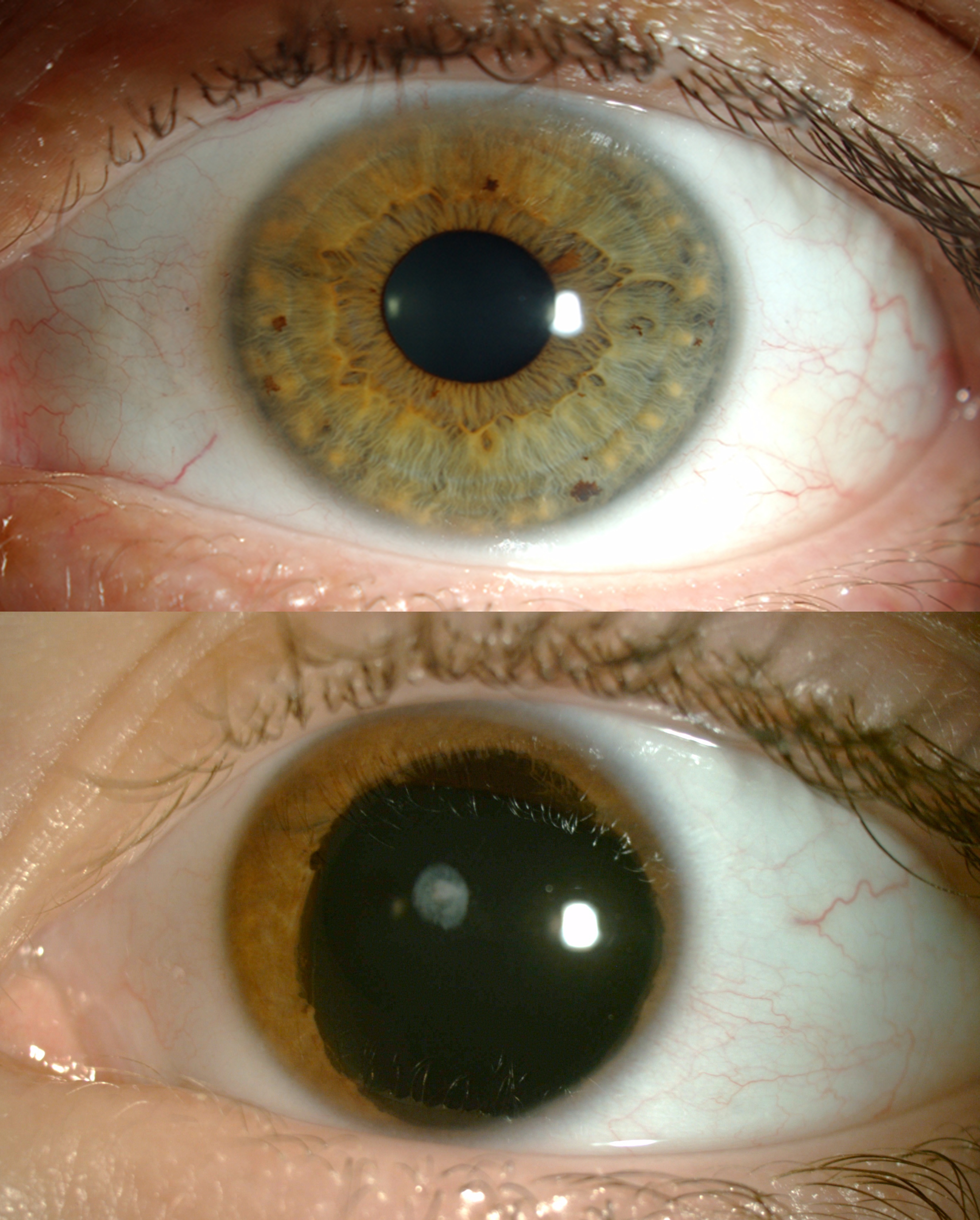
Aniridia is caused by the presence of a “nonsense mutation” – an extra “stop sign” on the gene that interrupts production of a protein crucial for eye development. Aniridia patients don’t have an iris (the coloured ring around the pupil), and suffer many other eye abnormalities.

Ataluren is believed to have the power to override the extra stop sign, thus allowing the protein to be made. The UBC-VCH scientists initially thought the drug would work only in utero – giving it to a pregnant mother to prevent aniridia from ever arising in her foetus. But then they gave their specially formulated Ataluren eye drops, which they call START, to two-week-old mice with aniridia, and found that it actually reversed the damage they had been born with.

“We were amazed to see how malleable the eye is after birth,” said Cheryl Gregory-Evans, associate professor of ophthalmology and visual sciences and a neurobiologist at the Vancouver Coastal Health Research Institute. “This holds promise for treating other eye conditions caused by nonsense mutations, including some types of macular degeneration. And if it reverses damage in the eye, it raises the possibility of a cure for other congenital disorders. The challenge is getting it to the right place at the right time.”

Bad vision at birth, worse vision later: Aniridia is apparent at birth because of the missing iris. Toddlers with aniridia need eyeglasses to see, sunglasses or darkened contact lenses to protect their eyes from overexposure to light, and cannot read small text. Their eyes are continually moving, making it difficult for them to focus, and have higher internal pressure (glaucoma), which damages the optic nerve as they get older. They are also prone to corneal damage in their teens and early adulthood. Eventually, most people with aniridia are considered legally blind, and must resort to Braille or expensive electronic aids to read.

An eye affected with aniridia on the bottom compared to an unaffected eye.

The plasticity of the eye: The reversal of tissue damage in young mice, published by the Journal of Clinical Investigation, fits with the fact that mammals’ eyes aren’t fully formed at birth. Human babies don’t discern colours until they are six months old, and their depth perception isn’t fully developed until the age of five.

Nonsense suppressor: Ataluren, made by the New Jersey-based PTC Therapeutics, is thought to be a “nonsense suppressor” – it silences the extra “stop codon” on the gene and allows a complete protein to be assembled. The drug is currently being tested as a treatment for cystic fibrosis and Duchenne muscular dystrophy, which are also caused by nonsense mutations.

Clinical trial: The forthcoming clinical trial, involving about 30 patients, will be led by Gregory-Evans in Vancouver, and is being supported by the Vision for Tomorrow Foundation, a US-based charity focused on aniridia and albinism (an absence of pigmentation in skin, hair and eyes that results in poor vision). If START is proven to be safe and effective, children with aniridia would use the drops twice a day for the rest of their lives. The drug would probably not reverse the condition in adults because their eyes would already be damaged beyond repair.

A gritty solution: Gregory-Evans’ first attempt at creating Ataluren eye drops proved unsuccessful. The drug didn’t dissolve, and thus irritated the mice’s eyes. So she turned to Kishor Wasan, a professor and associate dean in the Faculty of Pharmaceutical Sciences, who ground the drug into a very fine powder and combined it with a solution that adhered better to the eye.

A multidisciplinary team: Gregory-Evans also collaborated with her husband, Kevin Gregory-Evans, the Julia Levy BC Leadership Chair in Macular Research and an ophthalmologist at the VCH Eye Care Centre, who treats BC patients with aniridia. He administered the vision tests for the mice used in the study.

Funding from a UBC alumna: Cheryl Gregory-Evans’ position and this research were funded by the Sharon Stewart Testamentary Trust. Stewart, of Victoria, was born with aniridia, and at the time of her graduation from UBC in 1968, she only had five per cent of her vision. She died in 2008 and donated over $6 million to UBC for aniridia research.

Source; India Medical Times


Scientists identify potential drug to block AIDS

Scientists have identified an existing anti-inflammatory drug that in laboratory tests blocked the death of immune system’s cells which occurs as an HIV infection leads to AIDS.

Researchers are planning a Phase 2 clinical trial to determine if this drug or a similar drug can prevent HIV-infected people from developing AIDS and related conditions.

Scientists at the Gladstone Institutes identified the precise chain of molecular events in the human body that drives the death of most of the immune system’s CD4 T cells as an HIV infection leads to AIDS.

Two separate journal articles, published simultaneously in Nature and Science, detailed the research from the laboratory of Warner C Greene, who directs virology and immunology research at Gladstone, an independent biomedical-research nonprofit based in San Francisco.

His lab’s Science paper revealed how, during an HIV infection, a protein known as IFI16 senses fragments of HIV DNA in abortively infected immune cells.

This triggers the activation of the human enzyme caspase-1 and leads to pyroptosis, a fiery and highly inflammatory form of cell death.

As revealed in Nature paper, this repetitive cycle of abortive infection, cell death, inflammation and recruitment of additional CD4 T cells to the infection “hot zone” ultimately destroys the immune system and causes AIDS.

Nature paper further described laboratory tests in which an existing anti-inflammatory inhibits caspase-1, thereby preventing pyroptosis and breaking the cycle of cell death and inflammation.

“Gladstone has made two important discoveries, first by showing how the body’s own immune response to HIV causes CD4 T cell death via a pathway triggering inflammation, and secondly by identifying the host DNA sensor that detects the viral DNA and triggers this death response,” said Robert F Siliciano, a professor of medicine at Johns Hopkins University, and a Howard Hughes Medical Institute investigator.

Once the scientists discovered how CD4 T cells die they began to investigate how the body senses the fragments of HIV’s DNA in the first place, before alerting the enzyme caspase-1 to launch an immune response in the CD4 T cells.

They also identified that reducing the activity of a protein known as IFI16 inhibited pyroptosis.

“This identified IFI16 as the DNA sensor, which then sends signals to caspase-1 and triggers pyroptosis,” said Kathryn M Monroe, the Science paper’s other lead author, who completed the research while a postdoctoral fellow at Gladstone

Source: Times of India


Indian doctor awarded by American Society of Haematology

unnamed

Dr Praveen Kaudlay, a clinical haematology specialist registrar based in the UK, has been awarded with Abstract Achievement Award by the American Society of Haematology for his work as a first author on bone marrow failure condition called as paroxysmal nocturnal haemoglobinuria.


The award was presented, along with the paper presentation, at the 55th annual American Society of Haematology conference held in New Orleans, US from December 7 to 10.

Each year, the American Society of Haematology, offers merit-based Abstract Achievement Awards to select individuals to acknowledge the accomplishments of haematologists-in-training. This year’s Abstract Achievement Awards recognize undergraduate students, medical students, graduate students, resident physicians, and post-doctoral fellows who are both first author and presenter of an abstract.

Discussing about his paper with India Medical Times, Dr Kaudlay said, “The work was on bone marrow failure condition called as paroxysmal nocturnal haemoglobinuria. We looked at polymorphism in a complement molecule as these patients despite being treated with a drug, Eculizumab, tend to have haemolysis (blood getting destroyed inside the circulation). The study also included testing a molecule that blocks complement C3 in-vitro. The main investigator for this work is Prof Peter Hillmen, based at St James University Hospital at Leeds. I am the first author for the paper. Nearly 3,000 abstracts are submitted from around the world in this high profile conference attended by more than 20,000 delegates from all parts of the world.”


A graduate from Bangalore Medical College, Dr Kaudlay holds an interest in blood disorders and bone marrow failure syndromes and stem cell transplantation, which are quite common in India. He finished his postgraduation in General Medicine from India before going to the UK in 2003.

Talking about his future plans Dr Kaudlay said, “My future plan is to take research work that is of direct interest to India in blood disorders and bone marrow transplantation as Indians have different genetics compared to the Caucasians where most of the research are done. We have a large burden of thalassemia and sickle cell disease whose management needs to be improved. Bone marrow cancers are now being detected more than before needing novel chemotherapy drugs and stem cell transplantation. Indigenous research with Indian subjects needs to be pursued more to have a strong database to apply for treatment modalities.”

“My personal ambition is to develop a bone marrow donation registry which is as robust as in the West as patients of the Indian origin both within and outside India are struggling to find suitable match as lifesaving one marrow transplantation for many malignancies. There is a dire need to work along to develop the registry involving Indian population. The specialty of Haematology needs to be developed further to attract international trials and research in India,” he added.

Source: India medical times:


Diabetes risk gene may be inherited from Neanderthals


A gene variant that increases the risk of diabetes in Latin Americans may have been inherited from Neanderthals, a new study has found.

The gene variant was detected in a large genome-wide association study (GWAS) of more than 8,000 Mexicans and other Latin Americans.

People who carry the higher risk version of the gene are 25 per cent more likely to have diabetes than those who do not, and people who inherited copies from both parents are 50 per cent more likely to have diabetes, researchers found.

The higher risk form of the gene – named SLC16A11 – has been found in up to half of people with recent Native American ancestry, including Latin Americans, ‘BBC News’ reported.

The variant is found in about 20 per cent of East Asians and is rare in populations from Europe and Africa.

The elevated frequency of this risk gene in Latin Americans could account for as much as 20 per cent of the populations’ increased prevalence of type 2 diabetes – the origins of which are not well understood.

“To date, genetic studies have largely used samples from people of European or Asian ancestry, which makes it possible to miss culprit genes that are altered at different frequencies in other populations,” said co-corresponding author Jose Florez, a Broad associate member, an associate professor of medicine at Harvard Medical School and an Assistant Physician in the Diabetes Unit and the Center for Human Genetic Research at the Massachusetts General Hospital.

“By expanding our search to include samples from Mexico and Latin America, we’ve found one of the strongest genetic risk factors discovered to date, which could illuminate new pathways to target with drugs and a deeper understanding of the disease,” Florez said.

In the study published in the journal Nature, researchers conducted genomic analyses, in collaboration with Svante Paabo of the Max Planck Institute for Evolutionary Anthropology, and discovered that the SLC16A11 sequence associated with risk of type 2 diabetes is found in a newly sequenced Neanderthal genome.

Source: Indian Express


Inexpensive hearing devices bring gift of sound to less fortunate

It’s said the clacking of dominoes is the music of the Dominican Republic, but it’s been years since the game sounded so good to retiree Fernando Velverde Baez, who lost his hearing because of old age.

“The difference is that you hear the sound even of the air, and the breeze,” said Baez. “I hear a little bird, that little bird, that just chirped.”

Baez can hear the birds again because of Stavros Basseas, and a remarkable device from his company Sound World Solutions.

Basseas used two technologies, common even in the poorest countries, a smartphone that works with a personal sound amplifier in the ear, connected by Bluetooth. An app lets users raise or lower the volume, and adjust treble and bass.

If you are someplace that doesn’t even have cell phone service, you don’t need it, according to Basseas.

At up to $4,000, the tiny, high-tech hearing aids Americans can buy are too expensive for most of the estimated 365 million people worldwide with severe hearing loss.

Only seven million hearing aids were sold last year, and 85 percent of them ended up in American and European ears.

Basseas said his goal has been to “give people solutions in developing countries that don’t exist.”
And give them a price low enough that people can afford it — between $100 and $300.

The company plans to introduce the device in Africa and South America in the coming months. There may be profits down the road, but the goal for the moment is about helping.

Baez doesn’t care if his hearing aid is a little bulky or looks unfashionable, as long as he can now hear his wife say one thing. He said, “She would say, ‘I love you,’ and I wouldn’t be able to hear well. Now she can whisper it to me, and I’ll hear her.”

Source: one news page


Shock Therapy Can ‘Erase’ Bad Memories, Brain Researchers Show

In the film Eternal Sunshine of the Spotless Mind, unhappy lovers undergo an experimental brain treatment to erase all memories of each other from their minds. No such fix exists for real-life couples, but researchers report today in Nature Neuroscience that a targeted medical intervention helps to reduce specific negative memories in patients who are depressed.

“This is one time I would say that science is better than art,” says Karim Nader, a neuroscientist at McGill University in Montreal, Canada, who was not involved in the research. “It’s a very clever study.”

The technique, called electroconvulsive (ECT) or electroshock therapy, induces seizures by passing current into the brain through electrode pads placed on the scalp. Despite its sometimes negative reputation, ECT is an effective last-resort treatment for severe depression, and is used today in combination with anaesthesia and muscle relaxants.

Marijn Kroes, a neuroscientist at Radboud University Nijmegen in the Netherlands, and his colleagues found that by strategically timing ECT bursts, they could target and disrupt patients’ memory of a disturbing episode.

A matter of time
The strategy relies on a theory called memory reconsolidation, which proposes that memories are taken out of ‘mental storage’ each time they are accessed and ‘re-written’ over time back onto the brain’s circuits. Results from animal studies and limited evidence in humans suggest that during reconsolidation, memories are vulnerable to alteration or even erasure.

Kroes and his team tested this idea in 42 patients who had been prescribed ECT for severe clinical depression. In an initial session, the researchers showed two disturbing slide-show narratives: one depicting a car accident, and the other a physical assault.

The team later prompted patients to recall only one of the stories by replaying part of that slide show. Immediately afterwards, when the reactivated memory is thought to be vulnerable, the patients received electroconvulsive therapy.

One day later, when given a multiple-choice memory test, patients were significantly worse at remembering details from the reactivated story, performing near chance. Patients’ memory of the other story, however, remained largely unscathed. But when researchers administered the memory test 90 minutes after treatment, patients showed no differences in their ability to recall the two stories. This suggests that the therapy blocked the time-dependent process of reconsolidation, rather than causing sudden memory loss.

“This provides very strong and compelling evidence that memories in the human brain undergo reconsolidation, and that a window of opportunity exists to treat bad memories,” says Daniela Schiller, a neuroscientist at Mount Sinai Hospital in New York who also studies memory reconsolidation.

Thinking ahead
Schiller says more work is needed to establish how long the ECT effects last, and whether the technique works as effectively on older or more complex memories from real-life experiences, says Schiller.

Kroes adds that ECT may not be the best option for most patients, but says that these results could guide the development of less invasive interventions that target memory reconsolidation. Eventually, he says, the idea could be extended to memories involved in post-traumatic stress disorder, addiction and obsessive-compulsive disorder.

“The ability to permanently alter these types of memories might lead to novel, better treatments,” says Kroes.

Source: huffington post