MIT engineers develop paper diagnostic for cancer

Cancer rates in developing nations have climbed sharply in recent years, and now account for 70 per cent of cancer mortality worldwide. Early detection has been proven to improve outcomes, but screening approaches such as mammograms and colonoscopy, used in the developed world, are too costly to be implemented in settings with little medical infrastructure.

To address this gap, Massachusetts Institute of Technology (MIT) engineers have developed a simple, cheap, paper test that could improve diagnosis rates and help people get treated earlier. The diagnostic, which works much like a pregnancy test, could reveal within minutes, based on a urine sample, whether a person has cancer. This approach has helped detect infectious diseases, and the new technology allows noncommunicable diseases to be detected using the same strategy.

The technology, developed by MIT professor and Howard Hughes Medical Institute investigator Sangeeta Bhatia, relies on nanoparticles that interact with tumour proteins called proteases, each of which can trigger release of hundreds of biomarkers that are then easily detectable in a patient’s urine.

“When we invented this new class of synthetic biomarker, we used a highly specialized instrument to do the analysis,” says Bhatia, the John and Dorothy Wilson Professor of Health Sciences and Technology and Electrical Engineering and Computer Science. “For the developing world, we thought it would be exciting to adapt it instead to a paper test that could be performed on unprocessed samples in a rural setting, without the need for any specialized equipment. The simple readout could even be transmitted to a remote caregiver by a picture on a mobile phone.”

Bhatia, who is also a member of MIT’s Koch Institute for Integrative Cancer Research and Institute for Medical Engineering and Science, is the senior author of a paper describing the particles in the Proceedings of the National Academy of Sciences.

In 2012, Bhatia and colleagues introduced the concept of a synthetic biomarker technology to amplify signals from tumour proteins that would be hard to detect on their own. These proteins, known as matrix metalloproteinases (MMPs), help cancer cells escape their original locations by cutting through proteins of the extracellular matrix, which normally holds cells in place.

The MIT nanoparticles are coated with peptides (short protein fragments) targeted by different MMPs. These particles congregate at tumour sites, where MMPs cleave hundreds of peptides, which accumulate in the kidneys and are excreted in the urine.

In the original version of the technology, these peptides were detected using an instrument called a mass spectrometer, which analyses the molecular makeup of a sample. However, these instruments are not readily available in the developing world, so the researchers adapted the particles so they could be analysed on paper, using an approach known as a lateral flow assay — the same technology used in pregnancy tests.

To create the test strips, the researchers first coated nitrocellulose paper with antibodies that can capture the peptides. Once the peptides are captured, they flow along the strip and are exposed to several invisible test lines made of other antibodies specific to different tags attached to the peptides. If one of these lines becomes visible, it means the target peptide is present in the sample. The technology can also easily be modified to detect multiple types of peptides released by different types or stages of disease.

In tests in mice, the researchers were able to accurately identify colon tumours, as well as blood clots. Bhatia says these tests represent the first step toward a diagnostic device that could someday be useful in human patients.

“This is a new idea — to create an excreted biomarker instead of relying on what the body gives you,” she says. “To prove this approach is really going to be a useful diagnostic, the next step is to test it in patient populations.”

To help make that happen, the research team recently won a grant from MIT’s Deshpande Centre for Technological Innovation to develop a business plan for a startup that could work on commercializing the technology and performing clinical trials.

Bhatia says the technology would likely first be applied to high-risk populations, such as people who have had cancer previously, or had a family member with the disease. Eventually, she would like to see it used for early detection throughout developing nations.

Such technology might also prove useful in the United States, and other countries where more advanced diagnostics are available, as a simple and inexpensive alternative to imaging. “I think it would be great to bring it back to this setting, where point-of-care, image-free cancer detection, whether it’s in your home or in a pharmacy clinic, could really be transformative,” Bhatia says.

With the current version of the technology, patients would first receive an injection of the nanoparticles, then urinate onto the paper test strip. To make the process more convenient, the researchers are now working on a nanoparticle formulation that could be implanted under the skin for longer-term monitoring.

The team is also working to identify signatures of MMPs that could be exploited as biomarkers for other types of cancer, as well as for tumours that have metastasized.

Source: India Medical Times


Vinegar helps fight drug-resistant TB

A new study has found that the active ingredient in vinegar, acetic acid, might be used as an inexpensive and non-toxic disinfectant against drug-resistant tuberculosis (TB) bacteria as well as other stubborn, disinfectant-resistant mycobacteria.

“Mycobacteria are known to cause tuberculosis and leprosy, but non-TB mycobacteria are common in the environment, even in tap water, and are resistant to commonly used disinfectants. When they contaminate the sites of surgery or cosmetic procedures, they cause serious infections. Innately resistant to most antibiotics, they require months of therapy and can leave deforming scars,” senior author on the study, Howard Takiff, from Venezuelan Institute of Scientific Investigation (IVIC) in Caracas, said.

While investigating the ability of non-TB mycobacteria to resist disinfectants and antibiotics, Takiff’s postdoctoral fellow, Claudia Cortesia stumbled upon vinegar’s ability to kill mycobacteria.

Testing a drug that needed to be dissolved in acetic acid, Cortesia found that the control, with acetic acid alone, killed the mycobacteria she wanted to study.

It was found that exposure to 6 percent acetic acid, just slightly more concentrated than supermarket vinegar, for 30 minutes, reduced the numbers of TB mycobacteria from around 100 million to undetectable levels.

The study was published in the journal of the American Society for Microbiology.

Source: Businesss standard


Body Shape Index is Better Predictor of Mortality

In 2012, Nir Krakauer, an assistant professor of civil engineering in City College of New York’s Grove School of Engineering, and his father, Jesse Krakauer, developed a new method to quantify the risk specifically associated with abdominal obesity.

A follow-up study, published in PLoS ONE, supports their contention that the technique, known as A Body Shape Index (ABSI), is a more effective predictor of mortality than Body Mass Index (BMI), the most common measure used to define obesity.

The team analyzed data for 7,011 adults, 18+, who participated in the first Health and Lifestyle Survey (HALS1), conducted in Great Britain in the mid-1980s, and a follow-up survey seven years later (HALS2). The sample was broadly representative of the British population in terms of region, employment status, national origin and age. They used National Health Service records through 2009 to identify deaths and cancer cases: 2,203 deaths were recorded among the sample population.

Then, they compared all-cause mortality from the HALS sample with ABSI and other variables, including BMI, waist circumference, waist – hip ratio and waist – height ratio.

The analysis found ABSI to be a strong indicator of mortality hazard among the HALS population. Death rates increased by a factor of 1.13 (95 percent confidence interval, 1.09–1.16) for each standard deviation increase in ABSI. Persons with ABSI in the top 20 percent were found to have death rates 61 percent than those with ABSI in the bottom 20 percent.

The results tracked closely with the earlier study, which used data from the National Health and Nutrition Examination Survey (NHANES), conducted in the U.S. between 1999 and 2004. This provides stronger evidence that ABSI is a valid indicator of the risk of premature death across different populations. Further, they showed that ABSI outperformed commonly used measures of abdominal obesity, including waist circumference, waist – hip ratio and waist – height ratio.

Also, because the data came from two surveys seven years apart, the researchers were able to assess the effect of change in ABSI on mortality. The found an increase in ABSI correlated with increased risk of death, and that the more recent ABSI measurement was a more reliable predictor. Noting this, the researchers contend that further investigation is warranted into whether lifestyle or other interventions could reduce ABSI and help people live longer.

Source: laboratory equipment


Life Saver: Women With Cancer Gene Should Remove Ovaries by 35

Men with certain genetic mutations that greatly raise their risk of breast and ovarian cancer can cut the risk by as much as 80 percent if they get their ovaries removed by age 35, a new study suggests.

It’s the first study to show just how much the operation can do to lower the risk of cancer, and it’s the first to put such a clear age on the benefits.

It’s a finding so striking that the researchers think ovary removal should become standard for anyone with so-called BRCA1 mutations.

“This really validates for those of us who take care of women who have a high-risk BRCA1 … gene, that removing the ovaries and fallopian tubes really does have a positive impact on that woman, reduces her risk of ovarian cancer tremendously and also improves her survival,” said Dr. Ursula Matulonis of the Dana Farber Cancer Institute in Boston, who was not involved in the study.

It’s not an easy decision. Having the ovaries out, an operation called an oophorectomy, throws a young woman into instant and permanent menopause. But the study, published in the journal of Clinical Oncology, suggests it is worth all the awful side-effects, which include a likely end to a woman’s childbearing years.

Many women with BRCA mutations already get their breasts removed long before any sign of cancer — actress Angelina Jolie was one of them, having her breasts removed at age 37.

Nicole Armstrong knew she was at risk because her grandmother died so young. “So I always thought, ‘OK, I have an increased risk. Instead of going for mammograms at 50, I have to go around 40,” the 28-year-old, who lives in Easton, Pa.
Source: NBC news


Healthy liver cells created in lab

In a path-breaking research, scientists have discovered a way to transform skin cells into mature, fully functioning liver cells that flourish on their own.

The technique could serve as an alternative for liver-failure patients who do not require full-organ replacement or who do not have access to a transplant owing to limited donor organ availability.

Researchers at Gladstone Institutes and University of California, San Francisco (UCSF) revealed a new cellular reprogramming method that transforms human skin cells into liver cells that are virtually indistinguishable from the cells that make up liver tissue.

“Earlier studies tried to reprogramme skin cells back into a stem cell-like state in order to then grow liver cells. However, generating these pluripotent stem cells, or iPS cells, and then transforming them into liver cells was not always resulting in complete transformation,” explained Sheng Ding, senior investigator at Gladstone Institutes.

“So we thought that, rather than taking these skin cells all the way back to a stem cell-like state, perhaps we could take them to an intermediate phase,” he added

Instead of taking the skin cells back to the beginning, the scientists took them only part way, creating endoderm-like cells.

Endoderm cells are cells that eventually mature into many of the body`s major organs – including the liver.

This step allowed them to generate a large reservoir of cells that could more readily be coaxed into becoming liver cells.

Next, the researchers discovered a set of genes and compounds that can transform these cells into functioning liver cells.

After just a few weeks, the team began to notice a transformation.

“The cells began to take on the shape of liver cells and even started to perform regular liver-cell functions,” said Milad Rezvani from University of California.

They transplanted these early-stage liver cells into the livers of mice.

Two months post-transplantation, the team noticed a boost in human liver protein levels in the mice.

Nine months later, cell growth had shown no signs of slowing down.

These results offer new hope for the millions of people suffering from, or at risk of developing, liver failure.

At present, the only option is a costly liver transplant.

The power of regenerative medicine already allows scientists to transform skin cells into cells that closely resemble heart cells, pancreas cells and even neurons, concluded the study that appeared in the journal Nature.

Source: zee news


Soon, more efficient vaccines for meningitis and pneumonia

Researchers have discovered the presence of a novel subtype of innate lymphoid cells in human spleen essential for the production of antibodies.

This work was done by the B cell Biology research group at IMIM (Institut Hospital del Mar d’Investigacions Mediques) in Barcelona, directed by Dr. Andrea Cerutti, ICREA research professor and leader in the field of B lymphocyte biology.

Innate lymphoid cells were recently described by the scientific community and represent the first line of immunological defence on our body surfaces, which are constantly exposed to bacteria, such as the intestine or skin.

Dr. Giuliana Magri, member of the research group of B Cell Biology at IMIM and first author in the paper, said that for the first time it has been described both their presence and function in human spleen. We have discovered how these cells regulate the innate immune response of a subset of splenic B lymphocytes that are responsible to fight against encapsulated bacteria, causative agents of meningitis or pneumonia.

This new finding improves our understanding on how the immune system protects us against infections.

This research has been published in the journal Nature Immunology.

Source: sify news


Dogs can detect emotion in human voices, study shows

As many dog owners already know, dogs can often seem tuned into their “person’s” emotions or mood. Now, a new study from Current Biology offers evidence that this is, indeed, the case.

The study found that “dogs are sensitive to cues of emotion in human voices,”

And just how did researchers come to this conclusion? Fox reports, “For their study, researchers trained 11 dogs to sit still in a functional magnetic resonance imaging (fMRI) scanner. The researchers than analyzed the brain activity of both dogs and humans as they listened to 200 different dog and human sounds, ranging from crying to playful barking and laughing.

“While the brains of both dogs and humans responded most strongly to noises produced by their own species, they processed emotionally-loaded sounds in similar ways,” Fox reports.

“Some differences were noted as well: Dogs responded more strongly to non-vocal noises, compared to humans.”

Researchers hope the study will lead to “a better understanding of why dogs are so in tune with their owner’s emotions.”

Source: Now trending


AIIMS doctors devise new technique for complex spine surgery

0

Doctors at AIIMS have devised a new technique for complex spine surgery involving the joint at the upper part of the neck that supports and articulates with the head.

“The procedure called ‘distraction, compression, extension and reduction (DCER)’ removes the compression over the spinal cord and also reduces the deformity through a single surgery performed only from the back of the neck,” said Prof P Sarat Chandra (Dept of Neurosurgery) who conceptualised the innovative surgery.

The surgery normally is arduous, requires two separate procedures performed at the same sitting or in two different sittings and is a full day job, explained Chandra.

It first involves drilling out the piece of bone compressing the spinal cord (operated through the mouth) followed by a second procedure performed from behind the nape of neck resulting in stabilising the head and neck using rod and screws.

Surgery in this area is quite complex, technically demanding and requires intensive training. However, this procedure reduces the time of surgery by 50 per cent and has also been shown to reduce the rate of complications and hospital stay significantly.

The complication involving this area (called technically as atlanto-axial dislocation and basilar invagination) results in the uppermost portion of the neck slipping from its articulation with the base of the Lead causing compression of the spinal cord.

If untreated, the patient develops weakness of all limbs, becomes incontinent, bed-ridden and finally succumbs to the disease, said Chandra.

“The technique currently does not require any special instrumentation and utilises the existing instruments to perform a 2-axis motion which reduces the deformity,” stated Prof BS Sharma (Dept of Neurosurgery) who also has been part of the study.

The concept of the technique is connection based on the law of levers, first described by Archimedes, Prof P Sarat Chandra said.

He further added that the technique will be of immense use for our patients as it does not require any additional cost, reduces the hospital stay and also the complication rate thus benefiting the patients and the hospital in the long run.

The initial results of this procedure has been published in various prestigious international and national journals and has been also presented in various conferences.

Source: Business standard


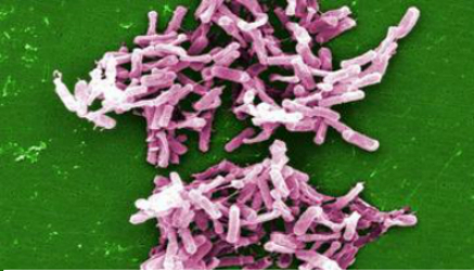
Fecal Transplant Regulations Are Too Strict, Researchers Say

c-difficile

Physicians use fecal transplants to treat certain intestinal infections, but the procedures recently came under strict regulations, with the Food and Drug Administration managing the transplants as though they were a drug treatment.

This regulation has made it harder for patients to receive fecal transplants, and in a new paper, some researchers are calling for the transplants to instead be regulated as a tissue, akin to blood donations.

The raw material for fecal transplants isn’t hard to come by, and so in the face of what some see as current over-regulation, an underground market for the transplants will likely spring up, the researchers argued today in the journal Nature.

At the same time, they said, more research is needed on the long-term effects of fecal transplants.

Regulating fecal transplants as a tissue may allow for better research on their possible uses in treatments, while protecting patients from harm, the researchers, from MIT and Brown University, wrote.

“I think regulating it as a tissue product would both provide access as needed and the research that could bring some pretty exciting new treatments on the scene,” said Mark B. Smith, an author of the article and a doctoral candidate at MIT.

Fecal transplants have been tested since the 1950s, and last year the first randomized controlled trial showed a strong benefit in helping patients with recurrent C. difficile, a bacterial infection that causes painful diarrhea, often following the use of antibiotics, and kills 14,000 people yearly in the United States.

But following the treatment’s success, some doctors began offering fecal transplants for other conditions as well — including those for which any potential benefit remains unproven. The FDA took action in 2013, regulating the treatment, but also granting an exemption for its continued use in patients who had C. difficile infections. This use would not require special permissions.

However, the result may be a case of both under- and over-regulation, today’s editorial argues. While medical societies have issued guidelines for using the treatment, there are no hard and fast rules for screening fecal matter, as there are with blood donations.

Source: Huffington Post


Saliva Test Predicts Risk of Severe Depression in Boys

High levels of stress hormone cortisol led to much greater likelihood of clinical depression diagnosis

If your teenage son is showing mild signs of depression, an experimental saliva test could determine if he’s at a risk for severe depression later in life.

In a study published in the journal Proceedings of the National Academies of Science, researchers examined the saliva of 1,800 teens between ages 12 and 19, and tracked the teens’ depression symptoms and mental illness diagnoses for up to three years later.

The test’s results were most pronounced for boys. Boys with mild symptoms of depression and high levels of cortisol–a stress hormone–were 14 times more likely to have a clinical depression diagnosis later on, compared with teens with lower levels of the hormone. For girls, high levels of cortisol put them at a four times greater risk for major depression later on.

Source: TIME