Japanese scientist withdraws ‘groundbreaking’ stem cell research

A Japanese scientist has asked for the findings of his groundbreaking study in stem cells to be withdrawn amid doubts over its quality.

According to Reuters news agency, Prof Teruhiko Wakayama of the University of Yamanashi told Japanese TV that when he was conducting the experiment, he believed that it was absolutely right, but many mistakes have emerged which has led him to withdraw the research paper, the BBC reported.

Several questions have been raised about the images used in the scientific report which claimed that dipping skin cells in acid could cheaply and quickly convert them into stem cells.

The original study, which was published in the journal Nature, had found that stem cells no longer needed to be taken from embryos or made by complicated and costly genetic tampering, and had been hailed as “remarkable” and as a “major scientific discovery”.

Source: Business Standard


Silk-based surgical implants could help heal broken bones

Researchers have developed surgical plates and screws which may not only offer improved bone remodeling following injury, but can also be absorbed by the body over time, eliminating the need for surgical removal of the devices.

Co-senior author Samuel Lin, MD, of the Division of Plastic and Reconstructive Surgery at BIDMC and Associate Professor of Surgery at Harvard Medical School, said that unlike metal, the composition of silk protein may be similar to bone composition, asserting that silk materials are extremely robust.

Lin and co-senior author and Tufts chair of biomedical engineering David Kaplan, PhD, used silk protein obtained from Bombyx mori (B. mori) silkworm cocoons to form the surgical plates and screws. Produced from the glands of the silkworm, the silk protein is folded in complex ways that give it unique properties of both exceptional strength and versatility.

To test the new devices, the investigators implanted a total of 28 silk-based screws in six laboratory rats. Insertion of screws was straightforward and assessments were then conducted at four weeks and eight weeks, post-implantation.

Lin said because the silk screws are inherently radiolucent [not seen on X-ray] it may be easier for the surgeon to see how the fracture is progressing during the post-op period, without the impediment of metal devices.

He said that having an effective system in which screws and plates ‘melt away’ once the fracture is healed may be of enormous benefit.

The findings have been published in the journal Nature Communications.

Source: zee news


Stem cell study sheds new light on disease formation

For the first time, researchers have shown that an essential biological process known as protein synthesis can be studied in adult stem cells.

The ground-breaking findings also demonstrate that the precise amount of protein produced by blood-forming stem cells is crucial to their function.

“This finding not only tells us something new about stem cell regulation but also opens up the ability to study differences in protein synthesis between many kinds of cells in the body,” said Sean Morrison, director of the children’s medical centre research institute at University of Toronto.

The discovery measures protein production, a process known as translation, and shows that protein synthesis is not only fundamental to how stem cells are regulated, but also is critical to their regenerative potential.

Different types of blood cells produce vastly different amounts of protein per hour, and stem cells in particular synthesise much less protein than any other blood-forming cells.

“This result suggests that blood-forming stem cells require a lower rate of protein synthesis as compared to other blood-forming cells,” Morrison added.

Researchers applied the findings to a mouse model with a genetic mutation in a component of the ribosome – the machinery that makes proteins – and the rate of protein production was reduced in stem cells by 30 percent.

The scientists also increased the rate of protein synthesis by deleting the tumour-suppressor gene ‘Pten’ in blood-forming stem cells.

In both instances, stem cell function was noticeably impaired.

Together, these observations demonstrate that blood-forming stem cells require a highly regulated rate of protein synthesis – such that increases or decreases in that rate impair stem cell function.

“Many people think of protein synthesis as a housekeeping function, in that it happens behind the scenes in all cells. The reality is that a lot of housekeeping functions are highly regulated,” explained Robert A J Signer, a post-doctoral research fellow in Morrison’s laboratory.

Many diseases, including degenerative diseases and certain types of cancers, are associated with mutations in the machinery that makes proteins.

Discoveries such as this raise the possibility that changes in protein synthesis are necessary for the development of those diseases, said the study published in the journal Nature.

Source: Times of India


New contraceptive ring aims to protect against both pregnancy and HIV

IntraVaginalRingNorthwestern

For decades, the condom has been the only form of contraceptive widely used to prevent both unplanned pregnancies and the transmission of HIV.

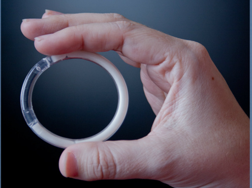
Now, researchers at Northwestern University have come up with a new option: An intravaginal ring that helps prevent pregnancy while simultaneously releasing low doses of an antiretroviral drug that reduces a woman’s risk of contracting both HIV and genital herpes.

Patrick Kiser, an associate professor of biomedical engineering and obstetrics and gynecology at Northwestern, devised the ring with the hopes that it would offer women more control over both disease and pregnancy prevention.

“The field of HIV prevention is really moving towards these long-acting drug delivery systems that require less user intervention, which is great because sex is episodic and exposure to [HIV] is episodic,” Kiser told FoxNews.com. “And because you don’t know when you’re going to be exposed, or even necessarily when you’re going to have sex, it’s better to…have protective measures on board at all time.”

Kiser and his team at Northwestern spent five years developing the two-inch ring, which releases doses of the contraceptive levonorgestrel and the common antiretroviral HIV medication tenofovir after being inserted in the vagina. Similarly to the NuvaRing, women can insert the device on their own. Women can then leave the ring in for up to 90 days, removing it briefly for cleaning if necessary.

Creating a device capable of releasing the proper doses of both the contraceptive and antiretroviral drugs posed a unique obstacle to researchers.

“The dose of contraceptive is very low – 10 micrograms per day, whereas with the antiviral drug we’re delivering is about 10 milligrams a day,” Kiser said. “That’s a thousand times different in terms of the amount being delivered for each drug and that was a real engineering challenge to develop a device that could achieve those extreme ranges of drug delivery.”

Eventually, they created a ring composed of three types of plastic tubing capable of releasing the appropriate doses of each medication contained within the device.

The medications used in the device both have a proven history of being both safe and effective. Levonorgestrel, which thickens a woman’s cervical mucus to prevent sperm from reaching the uterus, is widely used in popular forms of birth control like Mirena.

Source; Fox news


Doctors grow ears, noses using body fat stem cells

British scientists are aiming to grow ears and noses in a laboratory to transplant then into humans.

Scientists from Great Ormond Street Hospital and University College London have managed to use abdominal body fat and turn it into cartilage. It is now hoped that the technique could help patients who have been born with microtia, which means the ear fails to develop properly, or who have been in an accident.

At the moment, surgeons take cartilage from other parts of the body to treat children with facial defects. The painful procedure sees them shape the nose or ear, and implant it into the child

The new technique would mean that doctors ‘grow’ the organ separately using a tiny sample of fat from the child. Stem cells would be extracted and grown from it. Scientists would place an ear-shaped ‘nano-scaffold’ into the stem cell broth so that they take on the correct shape and structure.

This would then be placed beneath the skin. Although it would not help with hearing it would be biologically the same as the real thing, the Telegraph reports.  The breakthrough has been published in the journal Nanomedicine: Nanotechnology, Biology and Medicine.  Neil Bulstrode, consultant plastic surgeon at Great Ormond Street Hospital, co-authored the research.

He said: ‘It is such an exciting prospect. If we could produce a block of cartilage using stem cells and tissue engineering, this would be the holy grail for our field.’ Each year thousands of children are born with a congenital deformity called microtia, when the external ear is not fully developed. Many have an intact inner ear, but experience hearing loss due to the missing external structure.

But the research could also have implications for the future of other transplants, and could be used to create bone and other tissue. The report said the technique procedure could ‘help to improve stability, integration and functionality of engineered transplants while avoiding tissue rejection’.

Dr Patrizia Ferretti, the head of developmental biology at UCL, said it would be useful for children because it means there is no need for immune suppression.

She said: ‘At the moment we take cartilage out of the ribs which means a major additional surgical procedure that creates a permanent defect, as the rib cartilage does not regrow.
‘But with this technique you could seed the stem cells on to a mould of a healthy ear, or use 3D printing to make the ear shaped scaffold-containing cells that can then be turned into cartilage.’

Source: Daily mail


New sponge-like gel steers tooth formation

Inspired by this embryonic induction mechanism, Ingber and Basma Hashmi, a Ph.D. candidate at SEAS who is the lead author of the current paper, set out to develop a way to engineer artificial teeth by creating a tissue-friendly material that accomplishes the same goal. Specifically, they wanted a porous sponge-like gel that could be impregnated with mesenchymal cells, then, when implanted into the body, induced to shrink in 3D to physically compact the cells inside it.

To develop such a material, Ingber and Hashmi teamed up with researchers led by Joanna Aizenberg, Ph.D., a Wyss Institute Core Faculty member who leads the Institute’s Adaptive Materials Technologies platform. Aizenberg is the Amy Smith Berylson Professor of Materials Science at SEAS and Professor of Chemistry and Chemical Biology at Harvard University.

They chemically modified a special gel-forming polymer called PNIPAAm that scientists have used to deliver drugs to the body’s tissues. PNIPAAm gels have an unusual property: they contract abruptly when they warm.

But they do this at a lukewarm temperature, whereas the researchers wanted them to shrink specifically at 37°C — body temperature — so that they’d squeeze their contents as soon as they were injected into the body. Hashmi worked with Lauren Zarzar, Ph.D., a former SEAS graduate student who’s now a postdoctoral associate at Massachusetts Institute of Technology, for more than a year, modifying PNIPAAm and testing the resulting materials. Ultimately, they developed a polymer that forms a tissue-friendly gel with two key properties: cells stick to it, and it compresses abruptly when warmed to body temperature.

As an initial test, Hashmi implanted mesenchymal cells in the gel and warmed it in the lab. Sure enough, when the temperature reached 37°C, the gel shrank within 15 minutes, causing the cells inside the gel to round up, shrink, and pack tightly together.

“The reason that’s cool is that the cells are alive,” Hashmi said. “Usually when this happens, cells are dead or dying.”

Not only were they alive — they activated three genes that drive tooth formation.

To see if the shrinking gel also worked its magic in the body, Hashmi worked with Mammoto to load mesenchymal cells into the gel, then implant the gel beneath the mouse kidney capsule — a tissue that is well supplied with blood and often used for transplantation experiments.

The implanted cells not only expressed tooth-development genes — they laid down calcium and minerals, just as mesenchymal cells do in the body as they begin to form teeth.

“They were in full-throttle tooth-development mode,” Hashmi said.

In the embryo, mesenchymal cells can’t build teeth alone — they need to be combined with cells that form the epithelium. In the future, the scientists plan to test whether the shrinking gel can stimulate both tissues to generate an entire functional tooth.

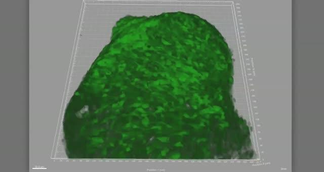
When the temperature rises to just below body temperature, this biocompatible gel shrinks dramatically within minutes, compressing tooth-precursor cells (green) enclosed within it.

As a new bioinspired, sponge-like gel shrinks, it squeezes cells (green) inside it, triggering them to shrink, round up, become denser, and begin to deposit the minerals that harden teeth.

Source; Science2.0


Microwaving tumors: New procedure knocks out kidney cancer without surgery

As a fight on cancer rages on, new record is creation it easier for doctors to mislay tumors though invasive surgery.

When Rory Kleinman, 42, sought medical courtesy for stomach issues in 2012, he had no suspicion that slight scans would exhibit a some-more critical problem.

“What happened was they were looking for something specific to do with my stomach, and by an MRI they afterwards saw something – a nodule on my liver – and so they had me do a successive MRI to check that,” Kleinman told FoxNews.com. “The nodule was fine, though in that second MRI they saw that there was a little mark that was on my kidney.”

That little mark on Kleinman’s kidney incited out to be a tumor.

“I only felt bombard shocked,” pronounced Kleinman. “I only never suspicion that we would have cancer during a immature age; if we was going to get it, we figured we would get it after in life.”

For many years, renal tumors compulsory prejudiced or sum dismissal of a kidney. Doctors would take a biopsy of a growth to see if it was cancer and afterwards confirm how many of a kidney to remove. But a new procession called x-ray ablation can be finished though surgery, and during a same time as a biopsy.

“Microwave ablation is a technique used to feverishness tumors,” Dr. Aaron Fischman, partner highbrow of radiology and medicine during Mount Sinai Hospital in New York City told FoxNews.com. “We’re means to indeed place a needle directly to a growth and kill it though indeed stealing it or creation an incision.”

Patients are put underneath unwavering sedation while a x-ray receiver is fed by a biopsy needle. After a square of a growth is private for testing, Fischman and his group use medical imaging to assistance place a tip of a receiver directly inside a tumor.

“The biggest advantage in my mind, and many of a patients will substantially tell you, that they don’t have to have surgery,” pronounced Fischman. “So we’re means to do this procession with no incision. We only put a needle directly into a kidney itself, and ablate it, so a liberation time is less, a snarl rate is theoretically reduction since a risk of draining is reduction though carrying a vital surgery.”

Microwave ablation is used to provide tumors in a liver, kidneys and lungs. Doctors during Mount Sinai have seen success rates of 90 to 95 percent in their patients who bear a procedure, Fischman said.

“Since this is a teenager procedure, a risks are minimal,” he said. “The many common thing that people can see is teenager draining or some pain during a site where a needle went in, and usually, this goes divided in a day or dual after a procedure.”

For Kleinman, a palliate of a procession has finished cancer a apart memory.

“Literally, we had a procession finished and a few days after we was behind during work – we unequivocally haven’t suspicion that many about it,” pronounced Kleinman. “I like that we don’t have to demeanour during a injure so that it reminds me that we had this procession done.”

Source: health medicine network


Will you die in 5 years? New ‘death test’ predicts

Scientists were astonished to find they could predict which healthy people are at most risk of death by studying four key biomarkers in the body

A ‘Death Test’ which predicts the chance of a healthy person dying from any medical condition in the next five years has been developed by scientists.

Researchers said they were ‘astonished’ to discover that a simple blood test could predict if a person was likely to die – even if they were not ill.

They found that the levels of four ‘biomarkers’ in the body, when taken together, indicated a general level of ‘frailty’.

People whose biomarkers were out of kilter were five times more likely to die with five years of the blood test.

“What is especially interesting is that these biomarkers reflect the risk for dying from very different types of diseases such as heart disease or cancer. They seem to be signs of a general frailty in the body,” said Dr. Johannes Kettunen of the Institute for Molecular Medicine in Finland.

“We believe that in the future these measures can be used to identify people who appear healthy but in fact have serious underlying illnesses and guide them to proper treatment.”

A biomarker is a biological molecule found in blood, body fluids, or tissues that may signal an abnormal process, a condition, or a disease.

The level of a particular biomarker may indicate a patient’s risk of disease, or likely response to a treatment. For example, cholesterol levels are measured to assess the risk of heart disease.

Most current biomarkers are used to test an individual’s risk of developing a specific condition. There are none that accurately assess whether a person is at risk of ill health generally, or likely to die soon from a disease.

Blood samples from over 17,000 generally healthy people were screened for more than a hundred different biomarkers and those people monitored over five years
In that time 684 people died of a range of illnesses and diseases, including cancer and cardiovascular disease. Scientists discovered that those people all had similar levels of four biomarkers.

Those were albumin, alpha-1-acid glycoprotein, citrate and the size of very-low-density lipoprotein particles which are linked to liver and kidney function, inflammation and infection, energy metabolism and vascular health.
One in five participants with the highest biomarker scores died within the first year of the study.

Estonian researchers made the initial link in a cohort of 9,842 people but were so sceptical about the results that they asked Finnish scientists to repeat the experiment on a further 7,503.

Research professor Markus Perola of the Institute for Health and Welfare, Finland said they were not expecting to be able to replicate the findings and were amazed when they were identical.

Prof Perola said: “It was a pretty amazing result. First of all we didn’t really believe it. It was astonishing that these biomarkers appeared to actually predict mortality independent of disease.

“These were all apparently healthy people but to our surprise it appears these biomarkers show an undetected frailty which people did not know they had.”

Researchers claim that in the future a test could flag up high-risk individuals in need of medical intervention who show no symptoms of any disease.

“If the findings are replicated then this test is surely something we will see becoming widespread,” added Prof Perola.

“But at moment there is ethical question. Would someone want to know their risk of dying if there is nothing we can do about it?”

Dr Kettunen added: “Next we aim to study whether some kind of connecting factor between these biomarkers can be identified.

Source: Telegraph


Finland researchers develop handheld camera for early detection of skin cancer

VTT Technical Research Centre of Finland has developed a lightweight, handheld, ultra-precision hyperspectral camera for the detection of skin cancers and their precursors.

From the surface of the skin, the camera recognises early stages of cancer that are invisible to the naked eye.

Collaborators in the pilot study are the University of Jyvaskyla, the Paijat-Hame Central Hospital and the Skin and Allergy Hospital of Helsinki University Central Hospital. The preliminary results are promising, say the researchers.

The hand-held, mobile hyperspectral camera images the skin region in two seconds. The large field of view (12 cm2) enables the detection of large skin areas at once.

In the pilot study, the camera has been used to detect the skin areas with field cancerization i.e. areas of multiple skin cancer precursors, actinic keratoses, for early treatment of the affected areas.

The hyperspectral camera has also been used to detect the borders of poorly delineated skin tumours, such as lentigo malignas, which are difficult to detect by the naked eye, in order to avoid the need for re-excisions.

Developed by VTT on the basis of the Fabry-Perot interferometer, the hyperspectral camera captures images in up to 70 narrow wavelengths, whereas a regular camera uses only three.

The spectral image generated is a three-dimensional cube built of numerous layers of greyscale images, each of which has been taken within a limited wavelength range.

A spectrum for each pixel of the spectral image is formed by the images within the cube. Different biological tissues can be identified by their reflected spectra in hyperspectral images.

Computational methods are used to interpret these images, in order to determine the position and size of the tumour to be treated. In the ongoing pilot study, all results are being verified by histopathological sampling.

Patents have been granted for the hyperspectral camera in the US and in Finland. Heikki Saari, principal scientist at VTT, is the inventor of this patented device.

Skin cancer rates have been growing exponentially, due to population ageing and UV damage caused by excessive exposure to sunlight.

The camera is owned by the University of Jyvaskyla. It can also be used for various applications of a more general nature, according to a statement by VTT.

Source: India Medical Times


Egypt claims miracle cure for HIV and Hepatitis

Two days ago, Egyptian media began big campaigns about a miraculous device (billed as ”Complete Cure”) invented by Egypt’s armed forces for treating HIV/AIDS and the Hepatitis C virus.

The news stories depicted the machine as a breakthrough, and a real miracle for completely curing patients of any of the two resistant diseases, giving hope for 18 million Egyptians with Hepatitis C and tens of thousands with HIV. (All this in no time at all — only 16 hours for the cure and one minute for detecting the disease.)

According to the Egyptian media, with the device there is no need to take a sample of the patient’s blood to detect the infection. Moreover, Egypt’s national TV channels ran a video that showed a physician making tests for an HIV patient using the device and telling him, “your tests are so great; you had HIV but now the disease vanished.”

The reports said that ”Complete Cure” is two machines in one: ”C Fast” (for treating Hepatitis C) and “I Fast” (for treating HIV). Furthermore, the reports confirmed that the military had been working for 22 years on the project and although ”C Fast” had been ready to go since 2006, the inventing team preferred to wait until it could test the effectiveness of the machine on HIV patients.

There’s even more. Major Dr. Ibrahim Abdel-Atti, leader of the machine-inventing team, stressed that Egypt will not export the machine to other countries so that it can be protected from international monopoly and the black market. He was quoted in the Al-Ahram newspaper as saying, “Marshal el-Sisi once said, ”we lagged behind and we should jump rather than walk so that we can compete with others”, and this is the first jump.”

The strange thing is, the media stresses that the device can also treat swine flu or H1N1, a disease that has taken many Egyptian lives over the past few weeks.

The media message is, of course, clear: We can depend only on Egypt’s armed forces; they’re the hope; they’re the people who can meet all our needs; they’re the men of impossible missions; they’re the best to lead Egypt in the coming years; they’re the best in the world; they’re big scholars and hard workers; they spare no effort to develop Egypt and improve the lives of its people.

They worked for 22 years for a machine that can make Egyptians happy. In short, the leader of Egypt must be a military man (in particular, Marshal Abdel-Fattah El-Sisi).

This kind of propaganda reminds us of old campaigns about the military’s invention of two inter-continental missiles: Zafer and Qaher, during Abdel-Nasser’s and Saddat’s era.

The two rockets were said to be able to reach to the depths of Israel. During that time, the media depicted this as if it was a nuclear bomb that could deter any threats to Egypt.

However, General Saad Ed-Dien el-Shazli mentioned in his diaries later that this was just propaganda and the two missiles were in fact ineffective and had no destructive power. El-Shazli stressed that he was shocked every time the media told lies about the two missiles and every time Saddat threatened America and Israel with using them.

In the current period, ordinary Egyptians are building mountainous hopes on such new devices and think that, with them, they can find light at the end of the tunnel.

The large majority of comments in the news describe the machine as a blessing and a gift from God, and many happy people have wondered aloud in the media about when the machine will be available in the hospitals. Many have been seen expressing their gratitude for the military and Marshal el-Sisi.

Source: the commentator