Mom of Newborn Twins Fights Rare Placenta Cancer

download

Jenna Hinman is fighting for her life in a medically-induced coma after she gave birth to healthy twins at 30 weeks pregnancy, only to discover she had a rare cancer of the placenta that has filled her lungs with tumors.

The 26-year-old mother is in “critical but stable” condition at Crouse Hospital in Syracuse, N.Y. The twin baby girls, Kinleigh and Azlynn are doing fine at 2 pounds, 9 ounces, and 3 pounds, 6 ounces, respectively.

“We’re hanging in there,” her husband, U.S. Army Sgt. Brandon Hinman, told ABCNews.com. “We are just taking it day to day, but we are starting to get some positive results and are pretty happy about that. The chemo is starting to have an effect.”

“The twins are doing well and don’t have breathing tubes anymore,” said Hinman, 30, who is stationed at Fort Drum. “Both are feeding and right where they need to be at 30 weeks.”
Crouse Hospital spokesman Bob Allen said her treatment was “a highly rare situation here, not just because of the pregnancy-related cancer, which is a big piece of it, but the fact that she is on ECMO technology.”

ECMO or extracorporeal membrane oxygenation is a therapy that uses a pump to circulate blood through an artificial lung back into the bloodstream. The most common conditions that may require ECMO are: heart malformations, severe air leak problems and severe pulmonary hypertension, according to the National Institutes of Health.

Jenna Hinman, a recreational therapist, began to go into labor on March 3 and was rushed to the emergency room at Good Samaritan Hospital in Watertown, N.Y., where the twins were delivered by emergency C-section. The same day, the twins were transported to Crouse Hospital in Syracuse where its Walter R.G. Baker Neonatal Intensive Care Unit is the only one in the in central New York designated as a regional perinatal center.

The new mother had only a quick visit with the babies before they were taken to the Crouse NICU. When Jenna Hinman began coughing up blood, she, too was transferred to Crouse.

“She was really in distress with breathing difficulties,” Crouse Hospital spokesman Cheryl Abrams told ABCNews.com. “It was a situation where the babies were in distress, too.”

At first doctors suspected pneumonia, but three days later, they diagnosed stage 3 choriocarcinoma.

Choriocarcinoma is a malignant form of gestational trophoblastic disease (GTD), tumors that involve abnormal growth of cells inside a woman’s uterus. This particular type affects only about 2 to 7 of every 100,000 pregnancies in the United States, according to the American Cancer Society.

Choriocarcinoma is much more likely than other types of GTD to grow quickly and spread to organs away from the uterus. About one-quarter of women who develop this disease miscarry.

Chief of Medicine for Crouse Hospital Dr. David Landsberg said stage 3 cancer had invaded Hinman’s lungs but had not gone to the brain. The cancer itself is “curable tumor,” he said.

“Placental tissue grows at a very rapid rate to support the fetus, which explains why the cancer is so aggressively metastatic,” he said. “It grows into the uterus and once it gets out, it’s looking for somewhere else to grow.”

Without ECMO, her condition would be “100 percent fatal,” said Landsberg. “The chemo will be the real cure. The ECMO is keeping her alive for the chemo to do its job.”

He said a cure was “on the edge of what is possible.”

Source: abc news


How aromatherapy can improve your health and how to use it effectively

The word “aromatherapy” probably conjures images of sweet-smelling massage oils, a bubble bath or candles made with synthetic scents. But while these products might smell nice, they don’t necessarily contain true healing oils. Authentic aromatherapy uses therapeutic plant extracts, which support the body in seeking balance and carry a variety of health benefits.

Plant extracts, also known as essential oils, are concentrates taken from roots, seeds, leaves or plant blossoms. Different oils, or combinations of oils, can be used in different ways. Some are used to promote physical healing, such as healing fungal infections, rashes or reducing pain and inflammation, while others are used as stress reducers or mood lifters.

Researchers are not entirely on clear how aromatherapy works. Some believe that sense of smell plays a major role, communicating with parts of the brain that serve to store emotions and memories and influence our physical, mental and emotional health. Others believe that it’s not about the aroma but the process that occurs in the body after their application and how the oils interact in the blood with hormones or enzymes.

Studies have shown that essential oils might have health benefits. As a result, aromatherapy is used in a variety of settings, from spas to hospitals, to treat various conditions. According to Cancer Research UK, it is a helpful complementary therapy for patients with cancer as well as other types of illness. Cancer patients noted improved quality of life, including reduced stress levels, better sleep and less feelings of sickness when undergoing aromatherapy treatments.

Generally, aromatherapy is used to improve mood, promote relaxation and relieve pain. In a study by the National Institutes of Health, participants who used aromatherapy had reduced stress levels during stressful situations. Relaxing and anxiety-reducing scents include lavender, vanilla, chamomile, frankincense and patchouli. Vibrant citrus aromas such as orange, lime and lemon have uplifting tendencies and are good choices to reduce depression. For muscle aches and headaches, chamomile, eucalyptus, ginger and lemon grass can all be used interchangeably.

There are several ways to enjoy the benefits of these healing oils. Diffusers and burners are a practical and effective way to reap the benefits of amomatherapy and are the most widely used methods of diffusing the healing properties of essential oils. Nebulizers process the oils into an almost gas-like state, making the oils easier to absorb and therefore more effective. Special oil-holding pendants can also be worn around the neck for portable benefits and adding a few drops to a bubble bath is a common relaxation technique. Aromatherapy massage is an excellent way to make the most of the benefits of essential oils – as your skin absorbs the oils you also breathe in their scent, all while reaping the stress-reducing benefits of massage.

Although essential oils are generally safe when used correctly, proper care should always be taken. Some oils can cause skin or allergic reactions and most oils need to be diluted before being applied directly to the skin. Since some essential oils can react with certain medications, or shouldn’t be used with certain health conditions, it is always best to get advice from a qualified aroma-therapist when choosing oils. Always remember to check that the oils you use are made from high quality plant ingredients and are not synthetic aromas claiming to provide aromatherapy benefits.

Source: fox news


Women Face Delays in Heart Attack Care: Study

Among young and middle-aged adults, men tend to receive faster hospital care than women for heart attacks and chest pains, a new study finds.

Anxiety appeared to be a key factor — women who appeared anxious upon admittance to the hospital tended to have delays in crucial care, the study authors found.

“Patients with anxiety who present to the emergency department with noncardiac chest pain tend to be women, and the prevalence of [heart attack or chest pains] is lower among young women than among young men,” the Canadian researchers said. “These findings suggest that [emergency-room staff] might initially dismiss a cardiac event among young women with anxiety.”

One heart expert wasn’t surprised by the findings.

“It has been shown in multiple trials that there are gender differences in the treatment of heart disease between men and women entering a hospital,” said Dr. Suzanne Steinbaum, director of women and heart disease at Lenox Hill Hospital in New York City.

“In younger adults, ages 18 to 55, this reality has also shown to be true,” she said. “When women enter a hospital, it is critical that this bias is eradicated.”

In the study, researchers looked at more than 1,100 adults aged 55 or younger treated for heart attack or chest pains — also called angina — at 24 hospitals in Canada, one in the United States and one in Switzerland. The median ages of the patients were 50 for women and 49 for men.

After arriving at the hospital, men underwent electrocardiograms (ECGs) within 15 minutes and clot-dissolving therapy within 21 minutes, compared with 28 minutes and 36 minutes, respectively, for women, the researchers said in the March 17 issue of the Canadian Medical Association Journal.

“Anxiety was associated with failure to meet the 10-minute benchmark for ECG in women but not in men,” said the researchers, led by Dr. Louise Pilote, a professor of medicine at McGill University in Montreal.

Digging deeper, the researchers found that patients with multiple heart attack risk factors and those with heart attack symptoms that were considered outside the norm also faced delays in care.

Steinbaum pointed out the incongruity of some of these findings.

“When analyzed, the women patients were sicker and were more likely to have diabetes, high blood pressure and a family history of heart disease,” she said. “With these multiple risk factors for heart disease, the likelihood of symptoms being heart-related are higher — yet they were not as expeditiously treated for a heart attack.”

“This delay in treatment is critical, especially in the setting of a heart attack, as death rates in patients who have multiple health problems is higher,” Steinbaum said.

Another heart specialist agreed.

“Since women often present with nontypical symptoms when having a heart attack, it is very important that physicians look at younger women, too, to make sure the symptoms they are having do not represent a developing heart attack,” said Dr. Lawrence Phillips, an assistant professor in the department of medicine at NYU Langone Medical Center in New York City.

“One of the most important take-home points from this study is the need to have an electrocardiogram early,” Phillips said. “An ECG is able to, in many cases, diagnose a heart attack as it occurs. By improving the rapidity of this test, we can start needed therapy earlier and, in doing so, save lives.”

Source: web md


Organ donation after cardiac death

organ

At only 32, Sarah Beth Therien suddenly became unconscious. She was rushed to hospital — and would never wake up. An unexpected heart arrhythmia had left her on life support. “A machine kept her heart pumping, but we knew she was gone,” said Emile Therien, her father.

After a week, Emile Therien and his wife, Beth Therien, made the difficult decision to withdraw life support.
Their daughter had always wanted to donate her organs, but she didn’t meet the brain death criteria required for donation. Her Ottawa family was determined to fulfill her final wish. In 2006, she became the first Canadian in nearly four decades to donate her organs after cardiac death — not brain death. And the decision didn’t go unnoticed.

Six years later in 2012, among the 540 deceased organ donors in Canada nearly 14 per cent donated after cardiac death. Cardiac death donation, also called non-heart-beating donation is now practised in Ontario, British Columbia, Alberta, Quebec, and Nova Scotia.

Canada joins other countries like the United Kingdom, United States, Spain, and the Netherlands, where non-heart-beating donation is more widespread.

Donation guidelines revised

Donation after cardiac death was the only method of deceased organ donation prior to the advent of brain death criteria in the 1960s — when the concept of someone being “brain dead” was first introduced. Because the brain dies before the heart, organs taken after brain death aren’t damaged from a lack of blood flow. As a result, donation after brain death replaced cardiac-death donation.

But over the last two decades, organ shortages, improved organ preservation, and public support led to the re-emergence of donation after cardiac death. In Canada, a national forum of transplant experts in 2005 led to the development of new guidelines that paved the way for this type of donation. And made it possible for Sarah Beth Therien to be a donor.

The potential impact is huge. Brain death accounts for only 1.5 per cent of in-hospital deaths. For the majority of patients with non-survivable illness, death occurs as a result of cardiac death after life support is removed.

Donation after cardiac death could increase the number of available organs by 10 to 30 per cent, according to experts. This could mean the difference between life and death for the nearly 4,500 Canadians currently waiting for a transplant, many of whom will die before getting organs.

The most common organs donated after cardiac death are kidneys, followed by livers, lungs, and pancreases.
Success depends on timing of organ removal

The success rate at which potential donors end up donating organs after cardiac death depends on the organs being removed — kidneys last longer than livers after the heart stops, for instance — how long it takes for the patient to actually die, and the person’s blood pressure and oxygen levels during the dying process, Shemie says.

And organs donated after cardiac death may not always work as well as those donated after brain death.

Higher rates of dysfunction have been seen in livers taken after cardiac death, says Dr. William Wall, director of the multi-organ transplant program at London Health Sciences Centre in Ontario. Kidneys donated after cardiac death have trouble working initially, but “one-year functioning is similar for kidneys taken after cardiac versus brain death,” Wall says.

For Emile and Beth Therien, pioneering the process meant a lot. Sarah Beth Therien donated two kidneys and two corneas, changing the lives of four Canadians. Each donor has the potential to save up to eight lives.

“Sarah was able to save other Canadians. Nothing could have made us happier,” Emile Therien says.

Source: CBC


Botox, Tattoo Removal, Now Possible in Kampala

tattoo-removal

But if you ever need to ditch those wrinkles for a smoother skin, or to remove that annoying tattoo or do hair replacement, you will definitely need it.

Internationally, aesthetic medicine is defined as “a branch of medicine that adheres to scientific-based procedures to enhance patients’ satisfaction with their physical appearance.”

In Uganda, aesthetic medical procedures are provided at Avane Cosmetic Clinic and medical spa along John Babiiha avenue, the International Medical Group and Kampala hospital. Below we explore the dos and don’ts of some of the common procedures.

Botox:

Botox is a protein, which when injected into a muscle causes it to become inactive or weakened. The most common uses for this treatment is smoothing of the facial wrinkles of the forehead, between the eyes and the corners of the eye and can also be used in the treatment of excessive sweating.

“Once these motor nerve endings are interrupted, the muscles cannot contract. Therefore, the motion that causes wrinkles in the skin will cease,” Dr Chiraag Kotecha, a dermatologist at Avane Cosmetic clinic and medical spa says. “Approximately three to 10 days after treatment, the skin above the motor muscles becomes smooth and nice.”

The effects of Botox last three to four months depending on the amount of protein injected and lifestyle activity of the patient. Do: For the first couple of years, Dr Kotecha recommends frequent treatments (every four to six months) for facial muscles to get used. Thereafter, treatments would be less frequent until a yearly maintenance is usually all that is required.

Don’t: Never let someone unprofessional administer the treatment on you as they may administer too much or too little of the protein.

Laser Hair removal:

Dr Pranav Pancholi, a visiting specialist in advanced non-surgical aesthetic medicine at Avane Cosmetic Clinic, says laser hair removal is more than just plucking out unwanted hairs. It is a medical procedure that reduces the amount of hair growth through a series of treatments, usually over a period of six to eight weeks and should be performed by a qualified physician.

The energy from the laser travels through the hair and destroys the follicle bump containing the cells responsible for hair re-growth,” explains Dr Pancholi. Patients seek hair removal for almost any body part, from the face, legs, arms, neck, back, chest and underarms. However:

Dos: Dr Pancholi advises one to undergo a skin test first to ensure that there will be no adverse side effects. He also advises one to do enough homework about the dermatologist’s qualifications and former clients. Don’t: Never undertake laser procedures if you are on light sensitive medication. Do not seek laser hair removal for gray hair because laser light only responds to dark pigmented hair follicles.

Tattoo removal surgery:

For the last one year, Mary (not real name) has been ridiculed at work for having a tattoo of a star on her left leg. Although she ignored the scoffing at first, it soon got to her and her promotion opportunities were limited because of her outward appearance.

“I was initially influenced by my peers to have the tattoo, but my hubby keeps telling me he does not like it,” she says, “Because it was affecting the attitude towards me at work, I have decided to come to Avane to remove it although it is quite expensive with every session costing me $50 (about Shs 130,000).”

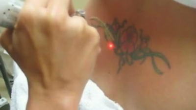
During treatment, special types of lasers known as Harmony XL lasers are used. Dr Kotecha says these are used break the pigment of the tattoo ink, dispersing it in the skin. Dos: Follow all the pre and post-treatment procedures carefully; ask your dermatologist if anything is unclear.

Don’t’s: “After your laser tattoo removal procedure, avoid any activity which submerges the treated area underwater for an extended period of time,” reads an article

Soaking the skin in water for an extended period of time, slows down the healing process and increases risk of infection.

Source; All Africa


Low-cost ultrasound machines may help reduce maternal mortality in rural areas

In one of the US Government’s Department of Health and Human Services’ reports for grants and guides, there has been a mention of the prerequisite of developing handheld ultrasound devices, especially for the detection of abscesses of lymph node enlargement, applicable to patients with HIV positive or negative. However, the need of ultrasound in medical services is huge as it is most popularly used for basic screening of high-risk pregnancies, cardiac, vascular, radiology, endocrinology and paediatric purposes. “It is most cost-effective and non-radiating machine that is used in any hospital across the globe. For a doctor who wants to have a quick insight of the reason behind the stomach pain, ultrasound is an immediate choice of diagnosis,” S Ganesh Prasad, director, ultrasound imaging, GE Healthcare, told India Medical Times.

Cost-Factor

According to Ganesh Prasad, ultrasound devices have existed in India for 20 years now but the significant market growth has happened in the last 10 years. “Prior to this, the ultrasound devices were available everywhere for the cost of 50,000 dollars (Rs 30 lakh) each but with the introduction of medical technology, low-cost devices have been produced and brought to various customers. Today, ultrasound machines are distributed for the cost ranging from Rs 2 lakh to Rs 70 lakh to various hospitals and medical experts.”

He further said, “The ultrasound machine worth Rs 2 lakh is a simple machine with which you can only do a simple scan to see womb or foetus, while the advanced ultrasound technology with higher cost provides you the real time assistance in witnessing the dynamic evolution of a foetus.”

GE Healthcare is doing a lot of research and development to improve the high quality ultrasound machine’s price form. “Our low-cost ultrasound devices range starts from Rs 4 to 5 lakh,” informed Ganesh Prasad.

“The cost of ultrasound machines depends on different gradations that the machine offers. Some are high-end machines of which prices go up to Rs 1 crore, mid-end machines cost around Rs 50 lakh and low-end machines are good enough for all the purposes, which cost up to Rs 12 to 15 lakh,” Dr Rakhee Gogoi, head of radiology, Paras Hospitals, Gurgaon, told India Medical Times while giving an estimate on the cost of the machines.

Quality Vs Low Cost

“We don’t believe in cheaper performance machine. We make products, which are good for the customers in terms of high technology. By the end of the year, we are planning to release a tablet ultrasound for the first time ever in the world. Every occupation could have it. For example, when a pregnant lady goes for a screening or check-ups, most of the time it’s only the physical exam that’s being done. Many a times, at the time of complication during delivery, there is no machine available for the doctors to see and learn the condition of foetus inside. The whole objective behind this technology is to make it relevant for you. We focus on target customer group and capability of technology, bringing it to a point of price that’s affordable,” said Ganesh Prasad.

GE’s low cost ultrasound devices come at the price point of Rs 5 lakh and products with a valid portfolio come with three years warranty. “We deploy current technology, not compromising in the quality at all levels. The whole idea behind offering three years warranty is to focus on quality as we understand that maintenance charges is a very good factor for the sustenance of ultrasound machines to a customer. We give the quality of a high-end machine but at the lower cost for people,” said Manoj Menon, communication leader, GE Healthcare India.

Penetrating Rural India

“Our villages do not have many ultrasound machines in rural areas. Ultrasound is very basic and important modality that can be used to detect any sort of pathology at a very early stage. Often, villagers come for the treatment when it’s too late for the problem till then the condition has gone out of hand. This is the reason why introduction and distribution of low-cost ultrasound machines will be really helpful in the rural areas,” said Dr Gogoi.

GE Healthcare is already planning to introduce low-cost ultrasound machines in the rural areas of India. “We have divided the market in tiers. Tier I is metro, tier II involves cities like Jaipur and tier III, IV and V have rural districts under them. We are doing good in tier I and tier II. In the tier III market, we have just stepped into. Besides producing the equipment, we are also creating a distribution network and affordability packages that has to be taken further,” said Menon.

Ganesh Prasad asserted, “Tier III is a fastest growing market with 22-23 per cent growth and that is going to be our area of focus. The reason why we have not penetrated as much in the tier III is because of the availability of the cost price for this kind of market. Our main effort has been to build a distribution network around tier II and the more challenging is going to be creating distribution around tier III.”

Improving Healthcare Situations

India has a high maternal mortality rate. “By proper screening during pregnancy, we can see improvement in the outcomes and avoid maternal or infant death during delivery. Ultrasound is the only device that can be used on a pregnant lady. Secondly, in rural areas, deliveries are still done at home which is intoxicating for a mother as well as for a child. With the introduction or availability of ultrasound machines in hospitals or nursing homes in tier III areas, pregnant women can be taken to hospitals for screenings. Some states have already taken the lead as Gujarat has improved its IMR rates and is moving towards greater institutional deliveries, while Tamil Nadu has also seen dramatic improvements in the reduction of infant mortality,” said Ganesh Prasad.

For Dr Jitendra Sharma, head, division of healthcare technology, National Health System Resource Centre, Ministry of Health and Family Welfare, the idea of handheld ultrasound devices clinically would be great and cost-effective. “It would be better than static ultrasound which comes for lakhs of rupees. What we are concerned about is social and regulatory aspect. Even with the static ultrasound machines in hospitals, we have not been practically able to control female abortions. If the hand held devices are sold in the market, let that market be rural or urban, how would female foeticide be controlled,” he told India Medical Times.

Controlling the Misuse for Sex Determination

Dr C V Bhirmanandham, vice president, Medical Council of India, told India Medical Times, “Why spend so much money when we can get good results from a low-cost ultrasound machine? But it should not be misused. The medical practitioners using the machines should be registered with a concerned authority and should be regularly monitored. Anyone caught misusing the machines for sex determination should be readily punished and their license should be cancelled.”

Dr Bhirmanandham also emphasised on the certification that the trainee must get from the concerned authority. “They have to follow the government of India policies and have to be ethical in their approach,” he added.

According to the Pre-Conception and Pre-Natal Diagnostic Techniques (PCPNDT) Act, 1994, the law prohibits any person from using ultrasound equipment for the detection of the sex of an unborn child. With an amendment in 2002 to the PCPNDT Act, the manufacturers and distributors of the ultrasound machines have to ensure the proper use of the equipment and are prohibited from selling, renting, permitting or authorizing the use of ultrasound machines for foetal sex determination. Manufacturers of ultrasound machines must also confirm that the customer has a valid PCPNDT certificate and they are also to provide the government with a quarterly report disclosing to whom the equipments have been sold.

“Hence, we don’t sell any of our ultrasound equipments to customers who are not registered under the PNDT Act. However, we believe that limiting the access of technology is more dangerous because we talk of saving lives and with the use of ultrasound technology, lives with different diagnosis can be saved. As per the government policies, we also continue to report to the government in every three months, disclosing about our dealings,” said Ganesh Prasad.

Source; India Medical Times


Doctors Use 3-D Printing To Help A Baby Breathe

Ever since the day Garrett Peterson was born, his parents have had to watch him suddenly just stop breathing.

“He could go from being totally fine to turning blue sometimes — not even kidding — in 30 seconds,” says Garrett’s mother, Natalie Peterson, 25, of Layton, Utah. “It was so fast. It was really scary.”

Garrett was born with a defective windpipe. His condition, known as tracheomalacia, left his trachea so weak the littlest thing makes it collapse, cutting off his ability to breathe.

“When he got upset, or even sometimes just with a diaper change, he would turn completely blue,” his mother says, “and that was terrifying.”So the Petersons contacted Dr. Glenn Green at the University of Michigan, who specializes in conditions like Garrett’s. He teamed up with Scott Hollister, a biomedical engineer who runs the university’s 3-D Printing Lab, to create a remarkable solution to Garrett’s problem — a device that will hold open Garrett’s windpipe until it’s strong enough to work on its own.

Instead of shooting ink onto a flat page to print words or pictures, 3-D printers use other material, such as plastic or metal, to create three-dimensional objects. “You build up layers until you have the complete 3-D structure,” Hollister says.

3-D printers have been used to build jewelry, art and even guns. But Hollister is using the technology to create medical devices. He uses a 3-D printer that melts particles of plastic dust with a laser. He has already built a jawbone for a patient in Italy and has helped another baby with a condition similar to Garrett’s. But Garrett is a lot of sicker and his condition is a lot more complicated.

“It’s just been issue after issue with breathing, and just trying to keep him breathing at all,” Jake Peterson, Garrett’s dad, says.

At 16 months old, Garrett had never been able to leave the hospital. Every time he stopped breathing, it was a mad rush to save him. And the doctors weren’t sure how much longer they could keep him alive.

“In some sense we were thrown directly into the fire,” Hollister says. “We characterized it as sort of a Hail Mary pass.” So they rushed Garrett from Salt Lake City to Ann Arbor on Jan. 18 and got to work.

First they took a CT scan of Garrett’s windpipe so they could make a 3-D replica of it. Next they used the 3-D printer to design and build a “splint.” It’s a small, white flexible tube tailored to fit around the weakest parts of Garrett’s windpipe.

“It’s like a protective shell that goes on the outside of the windpipe and it allows the windpipe to be tacked to the inside of that shell to open it up directly,” Green says.

But the device has not been approved by the Food and Drug Administration. So Green and Hollister had to convince the agency to give them an emergency waiver to try it. And they were running out of time. “His condition was critical. It was urgent and things needed to be done quickly. It was highly questionable whether he would survive and how long he would survive,” Green says.

Garrett’s parents knew they were taking a leap of faith. But they felt like they had to try. “We were just so excited for that glimmer of hope that this could be what would help Garrett get home,” Jake Peterson says.

Hollister and Green got the FDA’s approval and scheduled the surgery for Jan. 31. As soon as the surgeon, Dr. Richard Ohye, opened up Garrett’s chest, he and Green could see that Garrett’s windpipe had collapsed. One of his lungs was completely white.

“The only time I’d seen a white lung was in somebody that had died,” Green says. They quickly got to work, gingerly placing the first of two splints on one side of Garrett’s windpipe. It fit perfectly. So they got started on a second splint, which fit perfectly, too.

After more than eight hours, both splints were securely in place. Then came the most important moment: What would happen when they let air flow through Garrett’s windpipe into his lungs?

This time, Garrett’s windpipe stayed open, and his white lung turned pink. “That was just amazing to me,” Green says. “Here something that we’d worked on, that had been constructed just a week ago to match this defect. It had worked just the way we had hoped. I said, ‘This is going to change this boy’s life and his family’s life forever.’ ”

Garrett is 18 months old now and is still in the hospital, but in the weeks since the surgery, he has gotten stronger and stronger and needs less help breathing. His parents are ecstatic.

“He has been doing so good. He’s been smiling, and it’s crazy to be able to see him get really upset and not change colors,” Natalie Peterson says. “He’s being more interactive and more alert and reaching more for his toys. He’s just starting to be more like a normal child,” Jake Peterson adds. Garrett’s splint is designed to expand as he grows and eventually dissolve in his body as his own windpipe gets strong enough to work normally.

Green wants to save more babies this way, but it’s expensive to transport these extremely sick children across country. It has also been hard to convince insurance companies to pay for the trip.

“It is one of the most frustrating things that I’ve been through, knowing that there’s something that we have that can help and looking at all the roadblocks that are in place,” Green says. So he’s hoping to launch a formal study, which may enable him to try more splints to save more babies.

Green says this is the most exciting thing he has seen since medical school. “We’re talking about taking something like dust and converting it into body parts,” he says. “And we’re able to do things that were never possible before.” They’ve already started using 3-D printing to build more body parts, including ears and noses, by combining the plastic structure with human cells. Other scientists have gone even further, using 3-D printing to make blood vessels, skin and even primitive organs out of cells.

Source: npr


Outdoor exercise becoming popular

The gym appears to be giving way to outdoor exercises. Research shows that one in three women are shunning working out at the gym.

In an effort to workout in fresh air, 56 percent of health enthusiasts said they would definitely not be returning to the gym, femalefirst.co.uk reports.

The increasing popularity of functional workouts, boot camps and small group personal training sessions has fuelled the growth in the number of outdoor exercise options.

Being cheaper is also one of the main reasons women have decided to ditch the gym. But they also admitted that they feel more comfortable exercising in a less confined space with people watching.

Source: business standard


Study To Test ‘Chocolate Pills’ For Heart Health

It won’t be nearly as much fun as eating candy bars, but a big study is being launched to see if pills containing the nutrients in dark chocolate can help prevent heart attacks and strokes.

The pills are so packed with nutrients that you’d have to eat a gazillion candy bars to get the amount being tested in this study, which will enroll 18,000 men and women nationwide.

“People eat chocolate because they enjoy it,” not because they think it’s good for them, and the idea of the study is to see whether there are health benefits from chocolate’s ingredients minus the sugar and fat, said Dr. JoAnn Manson, preventive medicine chief at Harvard-affiliated Brigham and Women’s Hospital in Boston.

The study will be the first large test of cocoa flavanols, which in previous smaller studies improved blood pressure, cholesterol, the body’s use of insulin, artery health and other heart-related factors.

A second part of the study will test multivitamins to help prevent cancer. Earlier research suggested this benefit but involved just older, unusually healthy men. Researchers want to see if multivitamins lower cancer risk in a broader population.

The study will be sponsored by the National Heart, Lung and Blood Institute and Mars Inc., maker of M&M’s and Snickers bars. The candy company has patented a way to extract flavanols from cocoa in high concentration and put them in capsules. Mars and some other companies sell cocoa extract capsules, but with less active ingredient than those that will be tested in the study; candy contains even less.

“You’re not going to get these protective flavanols in most of the candy on the market. Cocoa flavanols are often destroyed by the processing,” said Manson, who will lead the study with Howard Sesso at Brigham and others at Fred Hutchinson Cancer Research Center in Seattle.

Participants will get dummy pills or two capsules a day of cocoa flavanols for four years, and neither they nor the study leaders will know who is taking what during the study. The flavanol capsules are coated and have no taste, said Manson, who tried them herself.

In the other part of the study, participants will get dummy pills or daily multivitamins containing a broad range of nutrients.

Participants will be recruited from existing studies, which saves money and lets the study proceed much more quickly, Manson said, although some additional people with a strong interest in the research may be allowed to enroll. The women will come from the Women’s Health Initiative study, the long-running research project best known for showing that menopause hormone pills might raise heart risks rather than lower them as had long been thought. Men will be recruited from other large studies.

Manson also is leading a government-funded study testing vitamin D pills in 26,000 men and women. Results are expected in three years.

People love vitamin supplements but “it’s important not to jump on the bandwagon” and take pills before they are rigorously tested, she warned.

“More is not necessarily better,” and research has shown surprising harm from some nutrients that once looked promising, she said.

Source: Huffington post


How to Unleash the Power of Garlic

This seasoning does more than kick up marinara. It stops sniffles, wards off UTIs, and may even help prevent cancer. Who knew garlic could have so many benefits?

Clear up UTIs
Cranberry juice isn’t the only natural way to fight annoying urinary tract infections. A diet rich in garlic can help, too, thanks to the bulb’s natural antimicrobial properties, says Amanda Ursell, author of The Complete Guide to Healing Foods. Garlic bread, anyone?

Crush cancer
The next time you’re cooking, mince the garlic ahead of time. Nutritionists at the American Institute for Cancer Research found that letting chopped or crushed garlic sit for 10 minutes before heating helps it retain a third more of its cancer-fighting sulfur compounds than if it were cooked immediately.

Fight itchy feet
Got athlete’s foot? Reach for garlic, a powerful antifungal, says Debra Rouse, a naturopathic physician in Denver. Just boil several cloves in hot water, cool to a comfortable temperature, pour into a large bowl, and soak your feet. (Boiled and uncrushed garlic won’t leave a smell.)

Combat a cold
At the first sign of the sniffles, try this get-well trick from Gowsala Sivam, PhD, of Bastyr University in Kenmore, Washington. Microwave 2 cloves unpeeled garlic for 25 seconds; let it cool, then peel off the skin and eat. Garlic’s sulfur compounds likely boost the immune system and help fight infection, Sivam says.

Let it bloom:
Plant garlic next to your rose bushes to naturally repel pests.

Source: health